


Impact of left ventricular geometry, expressed as interventricular septal and posterior wall thickness, on the outcome of acute ischemic stroke
- Special Hospital Sveti Sava, Belgrade, Serbia
- University of Kragujevac, Faculty of Medical Sciences, Kragujevac, Serbia
ABSTRACT
Introduction/Objective: Acute stroke represents a significant component of the overall health burden of the population. Parameters obtained by transthoracic echocardiography are well-established predictors of outcomes in cardiovascular diseases, while increasing evidence indicates their importance in predicting outcomes in neurovascular disorders.
This study aims to test the hypothesis that changes in left ventricular geometry, expressed as an increase in left ventricular wall thickness, represent a useful marker of poor outcome in patients with acute ischemic stroke.
Materials and methods: The methodology was based on the analysis of echocardiographic variables in 167 patients with acute ischemic stroke, together with variables of the modified Rankin Scale assessed at hospital discharge, used as an indicator of functional outcome.
Results: The study indicates a correlation between the thickness of both the interventricular septum and the posterior wall of the left ventricle, on the one hand, and the functional status of patients, on the other. The results confirmed the study hypothesis, demonstrating that an increase in interventricular septal thickness and/or posterior left ventricular wall thickness is associated with a higher probability of unfavorable outcome.
Conclusion: Both evaluated echocardiographic parameters showed positive predictive value in relation to functional outcomes.
INTRODUCTION
Stroke remains the second leading cause of mortality and, globally, represents the third most common combined cause of death and disability [1]. Furthermore, current projections indicate a continued increase in the incidence of this disease during the present decade, up to 2030, primarily in low-income countries and without significant differences with respect to sex or age [2]. Since 1990, the number of newly diagnosed stroke cases has been increasing in parallel with mortality, although age-standardized incidence rates have decreased over the same period [3]. A review of meta-analyses indicates that, in Europe, the incidence of stroke is approximately 191.9 per 100,000 person-years, with prevalence estimated at about 9.1% in men and 9.2% in women, and with a continuing upward trend [4]. Stroke represents the fourth leading cause of death in the United States [5].
Classical risk factors for atherosclerosis are also major risk factors for ischemic stroke; therefore, it is not surprising that in the vast majority of cases multiple cardiological comorbidities accompany stroke. This fact prompted the idea of evaluating basic parameters of left ventricular myocardial geometry obtained by transthoracic echocardiography – namely, the thickness of the interventricular septum (IVS) and of the left ventricular posterior wall (PW), as potential predictors of outcome in acute stroke.
The present study aims to assess the association between the thickness of the interventricular septum (IVS) and the posterior wall (PW) of the left ventricle, as “raw” and universally available bedside echocardiographic parameters that do not require more sophisticated computer programs, and the outcome of patients treated for ischemic stroke. This was done by testing the hypothesis that increased IVS thickness and left ventricular PW thickness are potential indicators of the outcome of this disease.
MATERIALS AND METHODS
Data were collected retrospectively from hospital medical records, after patient discharge, for a total of 167 patients treated for acute ischemic stroke in the Level III Intensive Care Unit of the Special Hospital Sveti Sava, between September 1, 2024 and September 1, 2025. In addition to the general demographic sample characteristics (sex and age of the participants), data were also collected regarding classical risk factors along with the examined parameters of transthoracic echocardiographic studies, namely the interventricular septum (IVS) thickness and the thickness of the left ventricular posterior wall (PW), measured in the parasternal longaxis view (PLAX).
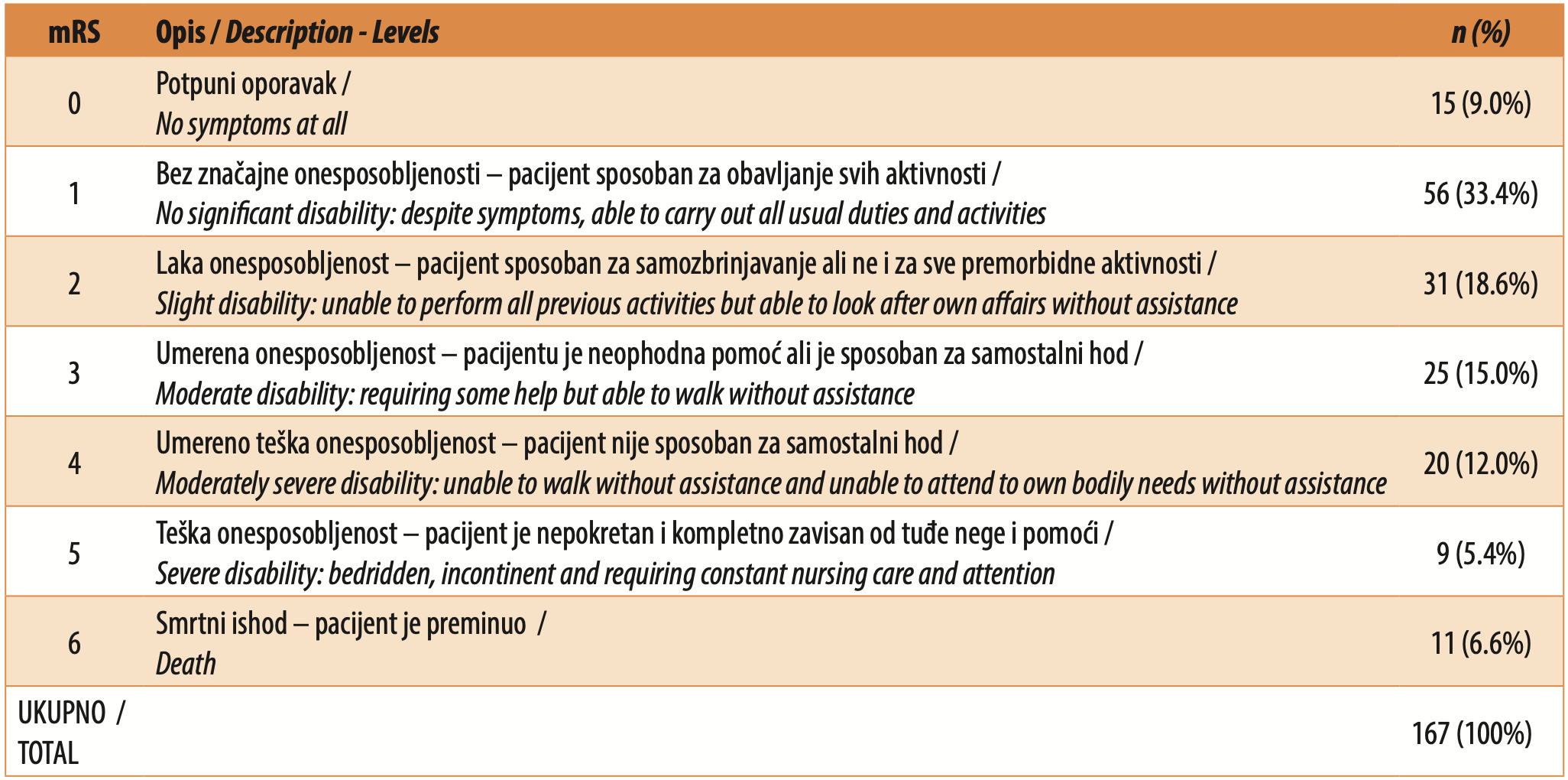
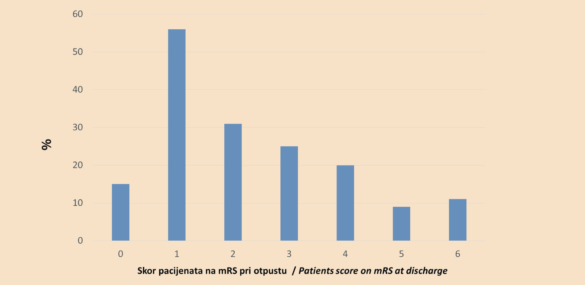
The achieved patient treatment outcomes were assessed using the modified Rankin Scale at hospital discharge and were taken to represent the indicator of disease outcome, as this scale is the most commonly used clinical tool for assessing the degree of disability after stroke. All ultrasound examinations were performed by an experienced cardiologist–echocardiographer within the first 72 hours after admission to the Intensive Care Unit, using a GE LOGIQ S8 device. Table 1 classifies the participants according to their score on the modified Rankin Scale (mRS), while Figure 1 is a graphical representation of the distribution of patients according to this characteristic.
The description of the data is presented in the form of absolute and relative frequencies (n, %) with the use of the arithmetic mean with standard deviation (mean ± SD) or the median (range, min-max), as dictated by the statistical form and distribution of the variables. The association between the degree of disease outcome and the examined echocardiographic statistical characteristics was evaluated using Spearman’s correlation coefficient. For testing the statistical significance of the working hypotheses, an alpha level of 0.05 was used. The IBM SPSS Statistics 24 (SPSS Inc., Chicago, IL, USA) software was used for statistical data analysis.
RESULTS
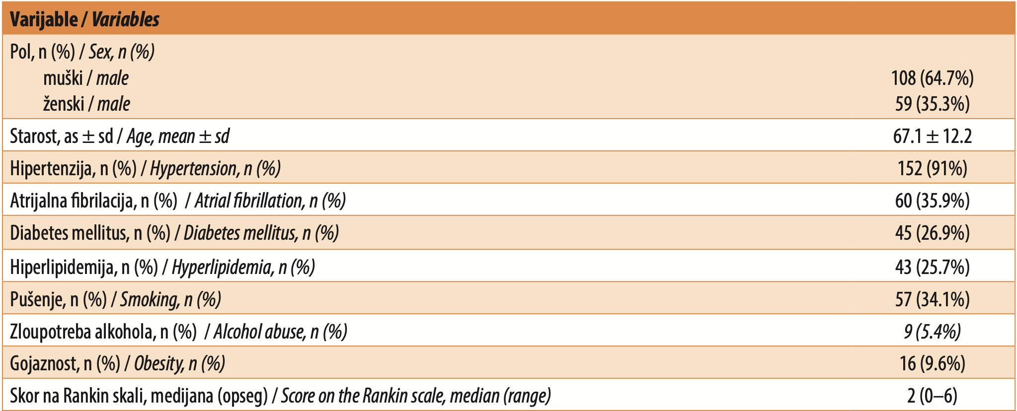
The analysis included a total of 167 patients treated for acute ischemic stroke. The average age of the sample was approximately 67 years (67.1 years with a standard deviation of 12.2 years), of whom 108 were male, and 59 were female. The oldest patient was 91 years old, while the youngest was 35 years old. The demographic characteristics of the sample and the distribution of risk factors are presented in Table 2. All analyzed subjects underwent a transthoracic echocardiographic study wherein the diameter of the interventricular septum (IVS) and of the left ventricular posterior wall (PW) were measured according to the standards of the American Society of Echocardiography (ASE) and the European Association of Cardiovascular Imaging (EACVI). The mean values for both variables with standard deviations are presented in Table 3.

The IVS diameter, defined in keeping with ASE/ EACVI recommendations as a universal and widely used marker of left ventricular geometry, was used as a determinant of disease outcome in the observed sample. Testing of treatment outcomes in relation to the IVS/PW diameter was carried out using non-parametric correlation between these variables and the final obtained score on the modified Rankin Scale.
Spearman’s correlation analysis showed a significant degree of correlation between disease outcome and the IVS diameter, with a positive correlation coefficient (rs = 0.91; p = 0.01; Table 4), indicating that poorer left ventricular geometry, expressed by an increase in IVS diameter, increases the degree of functional disability of patients after acute ischemic stroke.
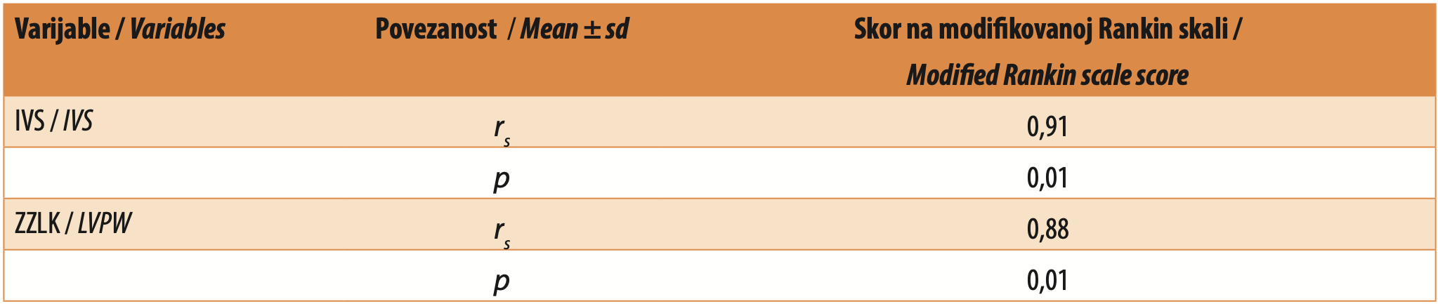
The diameter of the posterior left ventricular wall, defined in keeping with the same recommendations, was also tested as a potential variable for the prognosis of clinical outcome. Here also, a higher value of the variable significantly correlated with a higher score on the modified Rankin Scale (rs = 0.88; p = 0.01; Table 4), i.e., a larger diameter of the left ventricular posterior wall suggested a less favorable clinical course in patients with acute ischemic stroke.
DISCUSSION
The study included 167 patients of both sexes with acute ischemic stroke, with a predominance of men (64.7%). The overall mean age of the patients was approximately 67 years.
Among the collected, compared, and analyzed ultrasound characteristics of this sample, the diameter of the interventricular septum as well as the diameter of the posterior wall of the left ventricle, defined by the standards of the American Society of Echocardiography (ASE) and the European Association of Cardiovascular Imaging (EACVI) [6],[7], proved to be indicators of poor clinical outcome with a statistically relevant degree of positive association. Thus, an increase in the thickness of both the interventricular septum and the left ventricular posterior wall increases the degree of functional disability of patients after acute ischemic stroke.
In most modern studies, clearly defined parameters such as left ventricular mass or left ventricular relative wall thickness (LV RWT) are used as the basic markers of left ventricular geometry, which is certainly more appropriate because it better correlates prognostically with the outcomes of cardiovascular diseases [8–12]. However, such diagnostics requires more sophisticated echocardiographic software and is not applicable for bedside examinations such as those that are most feasible in acute stroke due to the condition of the patient. Another motive for working with basic data is the fact that raw data on IVS thickness have also been analyzed by other authors in patients with hypertension [13], which is traditionally considered the most clearly defined risk factor for stroke. It has been shown that increased interventricular septum thickness represents an independent and significant predictor of the future development of coronary disease and myocardial infarction. Furthermore, in the population of patients with atrial fibrillation included in the Atrial Fibrillation Follow-up Investigation of Rhythm Management (AFFIRM) study, left ventricular hypertrophy (LVH), defined as moderately or markedly increased interventricular septum thickness, proved to be a predictor of overall mortality and stroke, independent of classical clinical risk factors, while the definition of left ventricular hypertrophy based on IVS thickness had stronger and more consistent prognostic value compared with left ventricular mass alone [14].
In the general population, increased left ventricular mass assessed echocardiographically represents an independent and powerful predictor of adverse cardiovascular outcomes, including incident cardiovascular events and overall mortality, even after adjustment for traditional risk factors [15].
There are fewer studies dealing with the direct impact of interventricular septum thickness and/or left ventricular posterior wall thickness on the outcome of patients with acute ischemic stroke, but they do exist and are generally based on more advanced geometric data or on the assessment of left ventricular global systolic function. A study conducted by Adeoye et al. showed that interventricular septum thickness differed in patients with ischemic stroke, but it was not identified as an independent predictor of outcome after acute stroke [16]. We note that in this study the outcome was analyzed in a shorter, one-month period after ischemic stroke in a population that was younger than the population of our study, and that the research was conducted in Africa. Other authors have obtained data consistent with our research, namely that increased left ventricular relative wall thickness (LV RWT) was associated with higher mortality after ischemic stroke, suggesting that changes in the structure of the left ventricle, including wall thickening, have prognostic significance [17]. The association with our results is reflected in the fact that LV RWT, among other things, is calculated using the dimension of the posterior wall of the left ventricle.
Similar studies have also been conducted in patients with hemorrhagic stroke, who were not included in our research, but we mention them given that in this variant of stroke, according to the available literature, the thickness of the interventricular septum and the thickness of the left ventricular posterior wall did not show a significant association with disease outcome [18].
CONCLUSION
Both targeted ultrasound parameters show significant positive predictive value for the disease outcome. Namely, among the analyzed categories of patients, there is a statistically significant association between the diameters of both the interventricular septum and the left ventricular posterior wall and the achieved outcome of treatment of acute ischemic stroke, as assessed using the modified Rankin scale. The association is such that an increase in the thickness of the walls of the left ventricle favors a poorer functional outcome of stroke.
Stroke, as part of the spectrum of cardiovascular diseases, rarely occurs isolated from cardiological comorbidities, which themselves influence the manner of treatment of the underlying disease. Today, therapy is guided by modern algorithms, but the emphasis is still on individualization, which is dictated by the pathophysiology of all parallel comorbidities and other specific characteristics of each patient. Stroke is therefore commonly a multimorbid condition wherein there is interaction between pathological states, which creates the need for a multidisciplinary approach to the diagnosis, treatment, and assessment of patient prognosis.
LIST OF ABBREVIATIONS/ACRONYMS
IVS – interventricular septum
PW – posterior wall (of the left ventricle)
PLAX – parasternal long axis
ASE – American Society of Echocardiography
EACVI – European Association of Cardiovascular Imaging
LV RWT – left ventricular relative wall thickness
Informations

-
Keywords:
-
Received:
-
Revised:
-
Accepted:
-
Online first:
-
DOI:
-
Cite this article:

Dejan Munjiza
Special Hospital Sveti Sava
2 Nemanjina Street, 11000 Belgrade, Serbia
E-mail:
1. Feigin VL, Brainin M, Norrving B, Martins SO, Pandian J, Lindsay P, et al. World Stroke Organization: Global Stroke Fact Sheet 2025. Int J Stroke. 2025 Feb;20(2):132-44. doi: 10.1177/17474930241308142. [CROSSREF]
2. Pu L, Wang L, Zhang R, Zhao T, Jiang Y, Han L. Projected Global Trends in Ischemic Stroke Incidence, Deaths and Disability-Adjusted Life Years From 2020 to 2030. Stroke. 2023 May;54(5):1330-9. doi: 10.1161/STROKEAHA.122.040073. [CROSSREF] Erratum in: Stroke. 2024 Jan;55(1):e23. doi: 10.1161/STR.0000000000000452. [CROSSREF]
3. GBD 2019 Stroke Collaborators. Global, regional, and national burden of stroke and its risk factors, 1990-2019: a systematic analysis for the Global Burden of Disease Study 2019. Lancet Neurol. 2021 Oct;20(10):795-20. doi: 10.1016/S1474-4422(21)00252-0. [CROSSREF]
4. Soto A, Guillén-Grima F, Morales G, Muñoz S, Aguinaga-Ontoso I, Fuentes-Aspe R. [Prevalence and incidence of ictus in Europe: systematic review and meta-analysis]. An Sist Sanit Navar. 2022 Apr 28;45(1):e0979. Spanish. doi: 10.23938/ASSN.0979. [CROSSREF]
5. Imoisili OE, Chung A, Tong X, Hayes DK, Loustalot F. Prevalence of Stroke - Behavioral Risk Factor Surveillance System, United States, 2011-2022. MMWR Morb Mortal Wkly Rep. 2024 May 23;73(20):449-55. doi: 10.15585/mmwr. mm7320a1. [CROSSREF]
6. Lang RM, Badano LP, Mor-Avi V, Afilalo J, Armstrong A, Ernande L, et al. Recommendations for cardiac chamber quantification by echocardiography in adults: an update from the American Society of Echocardiography and the European Association of Cardiovascular Imaging. J Am Soc Echocardiogr. 2015 Jan;28(1):1-39.e14. doi: 10.1016/j.echo.2014.10.003. [CROSSREF]
7. Nagueh SF, Smiseth OA, Appleton CP, Byrd BF 3rd, Dokainish H, Edvardsen T, et al. Recommendations for the Evaluation of Left Ventricular Diastolic Function by Echocardiography: An Update from the American Society of Echocardiography and the European Association of Cardiovascular Imaging. J Am Soc Echocardiogr. 2016 Apr;29(4):277-314. doi: 10.1016/j.echo.2016.01.011. [CROSSREF]
8. Ghali JK, Liao Y, Cooper RS. Influence of left ventricular geometric patterns on prognosis in patients with or without coronary artery disease. J Am Coll Cardiol. 1998 Jun;31(7):1635-40. doi: 10.1016/s0735-1097(98)00131-4. [CROSSREF]
9. Yamaguchi S, Shimabukuro M, Abe M, Arakaki T, Arasaki O, Ueda S. Comparison of the prognostic values of three calculation methods for echocardiographic relative wall thickness in acute decompensated heart failure. Cardiovasc Ultrasound. 2019 Dec 3;17(1):30. doi: 10.1186/s12947-019-0179-6. [CROSSREF]
10. Zhang Y, Qiao S, Hao H, Li Q, Bao X, Wang K, et al. The predictive value of relative wall thickness on the prognosis of the patients with ST-segment elevation myocardial infarction. BMC Cardiovasc Disord. 2023 Jul 31;23(1):383. doi: 10.1186/s12872-023-03379-5. [CROSSREF]
11. Sun D, Schaff HV, Nishimura RA, Geske JB, Dearani JA, Ducharme MT, et al. Posterior Wall Thickness Associates With Survival Following Septal Myectomy for Obstructive Hypertrophic Cardiomyopathy. JACC Heart Fail. 2022 Nov;10(11):831-7. doi: 10.1016/j.jchf.2022.06.009. [CROSSREF]
12. Gardin JM. The Value of Left Ventricular Relative Wall Thickness in Predicting Ventricular Arrhythmia and Related Death. J Am Coll Cardiol. 2016 Jan 26;67(3):313-5. doi: 10.1016/j.jacc.2015.10.078. [CROSSREF]
13. Yang Y, Li Z, Guo X, Zhou Y, Chang Y, Yang H, et al. Interventricular Septum Thickness for the Prediction of Coronary Heart Disease and Myocardial Infarction in Hypertension Population: A Prospective Study. J Clin Med. 2022 Dec 1;11(23):7152. doi: 10.3390/jcm11237152. [CROSSREF]
14. Apostolakis S, Sullivan RM, Olshansky B, Lip GY. Left ventricular geometry and outcomes in patients with atrial fibrillation: the AFFIRM Trial. Int J Cardiol. 2014 Jan 1;170(3):303-8. doi: 10.1016/j.ijcard.2013.11.002. [CROSSREF]
15. Levy D, Garrison RJ, Savage DD, Kannel WB, Castelli WP. Prognostic implications of echocardiographically determined left ventricular mass in the Framingham Heart Study. N Engl J Med. 1990 May 31;322(22):1561-6. doi: 10.1056/NEJM199005313222203. [CROSSREF]
16. Adeoye AM, Ovbiagele B, Akinyemi JO, Ogah OS, Akinyemi R, Gebregziabher M, et al. Echocardiographic Abnormalities and Determinants of 1-Month Outcome of Stroke Among West Africans in the SIREN Study. J Am Heart Assoc. 2019 Jun 4;8(11):e010814. doi: 10.1161/JAHA.118.010814. [CROSSREF]
17. Park CS, Park JB, Kim Y, Yoon YE, Lee SP, Kim HK, et al. Left Ventricular Geometry Determines Prognosis and Reverse J-Shaped Relation Between Blood Pressure and Mortality in Ischemic Stroke Patients. JACC Cardiovasc Imaging. 2018 Mar;11(3):373-82. doi: 10.1016/j.jcmg.2017.02.015. [CROSSREF]
18. Tatar S, Kesriklioğlu S, Şalvarcı A, Yaşaroğlu MY, Tokgöz OS. The effect of aortic arch calcification on in-hospital mortality in patients with hemorrhagic stroke. Neurology Asia. 2025 Mar;30(1):19-25. doi: 10.54029/2025uyc. [CROSSREF]
- Table 1. Distribution of responses on the Modified Rankin scale (mRS)
- Figure 1. Relative frequency distribution of modified Rankin Scale categories
- Table 2. Demographic variables, risk factors and median score of disease outcome
- Table 3. Echocardiographic parameters
- Table 4. Spearman’s correlation coefficients and p-values