


Clinical characteristics and the treatment outcome in patients with acute lymphoblastic leukemia
- General Hospital Valjevo, Valjevo, Serbia
- Clinic for Hematology, University Clinical Center of Serbia, Belgrade, Serbia
- Faculty of Medicine, University of Belgrade, Belgrade, Serbia
ABSTRACT
Introduction: Acute lymphoblastic leukemia (ALL) is the malignant transformation and proliferation of lymphoid progenitor cells. The disease outcome is significantly influenced by cytogenetic and molecular characteristics, leukocyte levels, the percentage of blood and bone marrow blasts, infiltration of the CNS, and the modality of the applied therapy.
Aim: Determining clinical, immunophenotypic, and cytogenetic characteristics of patients with ALL; ascertaining remission rates, the presence of resistant forms, the rates of relapse and overall survival (OS); determining prognostic factors for patient survival.
Materials and methods: This five-year retrospective study included 74 patients. The study covered the period from November 2013 to October 2018. The influence of sex, age, immune subtype of ALL, leukocyte levels, percent of blood and bone marrow blasts, and cytotoxic-molecular characteristics on survival were analyzed.
Results: A total of 56 (75.0%) patients had B-cell ALL, while 18 (25.0%) patients had T-cell ALL. The average age was 49.5 years (20 - 77). Complete remission was achieved in 54 (72.9%) patients, 40 (71.4%) B-cell ALL patients and 14 (77.7%) T-cell ALL patients. Refractory leukemia was confirmed in 7 (9.4%) ALL patients, 5 (8.9%) B-cell ALL patients and 2 (11.1%) T-cell ALL patients. Mortality during induction was observed in 13 (17.56%) patients, 8 (14.2%) B-cell ALL patients and 5 (27.7%) T-cell ALL patients. Relapse occurred in 25 (46.3%) patients, 20 (47.5%) B-cell ALL patients and 5 (33.3%) T-cell ALL patients. Univariate analysis showed that an unfavorable outcome was associated with age ≥50 years (p < 0.001), COP chemotherapy (p < 0.001), non-transplant patients (p = 0.011), and infiltration of the CNS (p < 0.001).
Conclusion: Survival in ALL patients is significantly affected by patient age, modality of applied therapy, and infiltration of the CNS by disease.
INTRODUCTION
Acute lymphoblastic leukemia (ALL) is a heterogeneous disease characterized by the malignant transformation and proliferation of lymphoid progenitor cells in the bone marrow, peripheral blood, and other tissues [1].
The incidence of ALL in Europe is 1.28 per 100.000 individuals annually, with significant variations in relation to age [1]. The highest percentage of individuals affected by this disease, almost 80.0%, is registered in the pediatric population, whose survival rate is 90.0%, while the adult population has a significantly poorer prognosis [2].
According to immunological classification, there are two types of acute lymphoblastic leukemia: B-cell ALL and T-cell ALL. There are four subtypes within the B-cell type of ALL: pro B-ALL, common B-ALL, pre B-ALL, and immature B-ALL. The subtypes of T-cell ALL are classified as: pro T-ALL, pre T-ALL, cortical T-ALL, and mature T-ALL [3].
The most significant prognostic factor in acute lymphoblastic leukemia is the existence of cytogenetic chromosomal abnormalities. Numeric chromosomal aberrations (tertarploidy, hypodiploidy, and pseudodyploidiy) represent a poor prognostic factor, and the survival rate in these individuals is 25.0%. In persons with ALL, the most analyzed and best known structural chromosomal aberration is translocation t(9;22) (Philadelphia chromosome). This aberration is apparent in 20.0% - 30.0% of adult patients suffering from ALL. In addition to t(9;22), translocations t(8;14) and t(4;11) are also poor prognostic factors [4]. Besides cytogenetic abnormalities, the prognosis in acute lymphoblastic leukemia is also affected by sex (greater number of relapses in the male sex), age (poorer prognosis in adults older than 60 years), time necessary for achieving complete remission (CR), leukocyte count (>30×109 for B-cell ALL and >100x109 for T-cell ALL), and central nervous system (CNS) involvement [1],[5]. The presence of immature precursor T and B cells is also linked to a poor prognosis, which is why the presence of B cells which manifest CD19, CD22 and CD79a on their surface, as well as the presence of T cells which do not manifest CD1a, CD8 and CD5, are believed to be poor prognostic factors [6].
Although remission is achieved in almost 90.0% of the patients, after initial chemotherapy (CT), the cure rate is only 40.0% - 50.0%, due to relapse. Conventional chemotherapy regimens applied in patients with relapse or with the refractory form of B-cell ALL are linked to the survival rate of 31% to 44%, if administered in the early stage of relapse, and with a survival rate of 18.0% - 25.0%, if administered at a later stage of relapse or as a second-line CT treatment option [7].
Hematopoietic stem cell transplantation (HSCT) has proven to be very efficient in the treatment of high-risk patients suffering from ALL. Five-year survival in patients in whom minimal residual disease (MRD) after transplantation is not registered is 75.0%, while the survival rate in patients with a positive MRD status is 33.0% [8].
The aims of this study were to determine clinical, immunophenotypic, and cytogenetic characteristics of ALL patients, as well as to to ascertain complete remission rates, the presence of resistant forms, the rates of relapse, and overall survival (OS) in ALL patients. Also, the aim was to determine prognostic factors affecting ALL patient survival.
MATERIALS AND METHODS
The analyzed group included 74 ALL patients; 56 (75.0%) patients with B-cell ALL and 18 (25.0%) patients with T-cell ALL. The study is retrospective, and it encompasses a five-year period of patient follow-up, from November 2013 to October 2018. The diagnosis of the disease was established based on clinical presentation and on peripheral blood and bone marrow findings, in keeping with World Health Organization (WHO) recommendations [9],[10]. The following specific parameters were monitored in patients: sex, age, immunological subtype of acute lymphoblastic leukemia, leukocyte count, percentage of blasts in peripheral blood and bone marrow, molecular cytogenetic characteristics. The group of risk factors for acute lymphoblastic leukemia was determined according to the GMALL (German Multicenter Study Group for Adult ALL) risk factor group model and it included standard, high and very high level of risk [11].
Hematological diagnostics included:
- Morphological and cytochemical analysis of the bone marrow smear
Bone marrow smears were first dyed with the application of the May-Grünwald-Giemsa method, after which specific cytochemical staining techniques were applied (PAS, POX, SB, ANAE). Cytochemical staining makes it possible to determine the lineage of the blasts. At the same time, the cellularity, presence, number, and the maturation stage of individual hematopoietic cells, as well as their morphological characteristics were determined [9],[10].
- Immunophenotypization by means of flow cytometry
The samples of the test subjects were collected and processed according to the standard recommendations and protocols, with the application of the method of direct multicolor immunofluorescence [12].
- Classical cytogenetic analysis with the application of the method of HG strips
The patient’s karyotype, at the time of establishing diagnosis, was determined with the use of the G banding technique and analyzed in keeping with the International System for Human Cytogenetic Nomenclature [13].
- Molecular – genetic study
The following procedures were performed within molecular genetic studies: isolation of mononuclear cells from bone marrow aspirate, isolation of RNA from mononuclear cells, reverse transcription (RT), standardized RT-PCR (polymerase chain reaction) protocol for the detection of BCR-ABL fusion transcripts.
Treatment of ALL patients
ALL patients were treated with the Hyper-CVAD and COP chemotherapy protocols. The Hyper-CVAD protocol consists of even and odd treatment courses applied in patients aged ≤ 65 years, who had an ECOG (Eastern Cooperative Oncology Group) ≤ 2 and an HCT-CI (hematopoietic cell transplantation-comorbidity index) ≤ 3. The odd course of treatment includes the application of the following: cyclophosphamide, 300 mg/m2 IV, D1-3; mesna, 600 mg/m2 IV, D1–3; doxorubicin, 50 mg/m2 IV, D4; vincristine, 2 mg IV, D4 and D11; dexamethasone, IV 40 mg, D1–4 and D11–14. The composition of the even course of treatment is the following: methotrexate (MTX), 200 mg/m2 and 800 mg/m2 IV, D1 for 22h; and high doses of cytosar (Ara-C) 3 g/m2 /12 h, D2 and D3. The maintenance therapy over a period of two years included the application of: 6-mercaptopurine per os, 3x50 mg per day; MTX, IM 20 mg/m2 , once a week; prednisone, 200 mg a day, D1–5 in the month; vincristine, 2 mg IV, once a month. Patients with ALL received CNS prophylaxis. The COP protocol was applied in patients who had ECOG >2, HCT-CI >3, >65 years, and consisted of the following: cyclophosphamide amp, 750 mg/m2 IV, D1; oncovin, 2 mg IV, D1; and prednisone tbl, 100 mg p.o., D1- 5. Allogeneic hematopoietic stem cell transplantation (HSCT) was carried out in ALL patients who had a high or very high level of risk. Complete remission (CR) means ≤ 5.0% of blasts in the bone marrow, while failing to achieve full remission after two cycles of chemotherapy is considered to be refractory disease. Overall survival (OS) is defined as the time from diagnosis until death or the last follow-up date. Disease-free survival (DFS) is defined as the time from achieving complete remission until relapse or death. Early death (ED) is defined as death within 28 days of starting induction chemotherapy.
In the statistical processing of data, methods of descriptive statistics were applied. For analyzing the difference in frequency, the Chi-square test, i.e., the Fisher’s exact test was applied. For the analysis of the length of survival, the Kaplan-Meier method was used, while the logrank test was applied for comparing survival amongst the test groups. The univariate Cox proportional hazards regression model was used for identifying risk factors. All statistical tests were observed at the null-hypothesis significance level of p < 0.05. Statistical analysis was performed on a PC with the application of a licensed SPSS 15.0 statistical package.
RESULTS
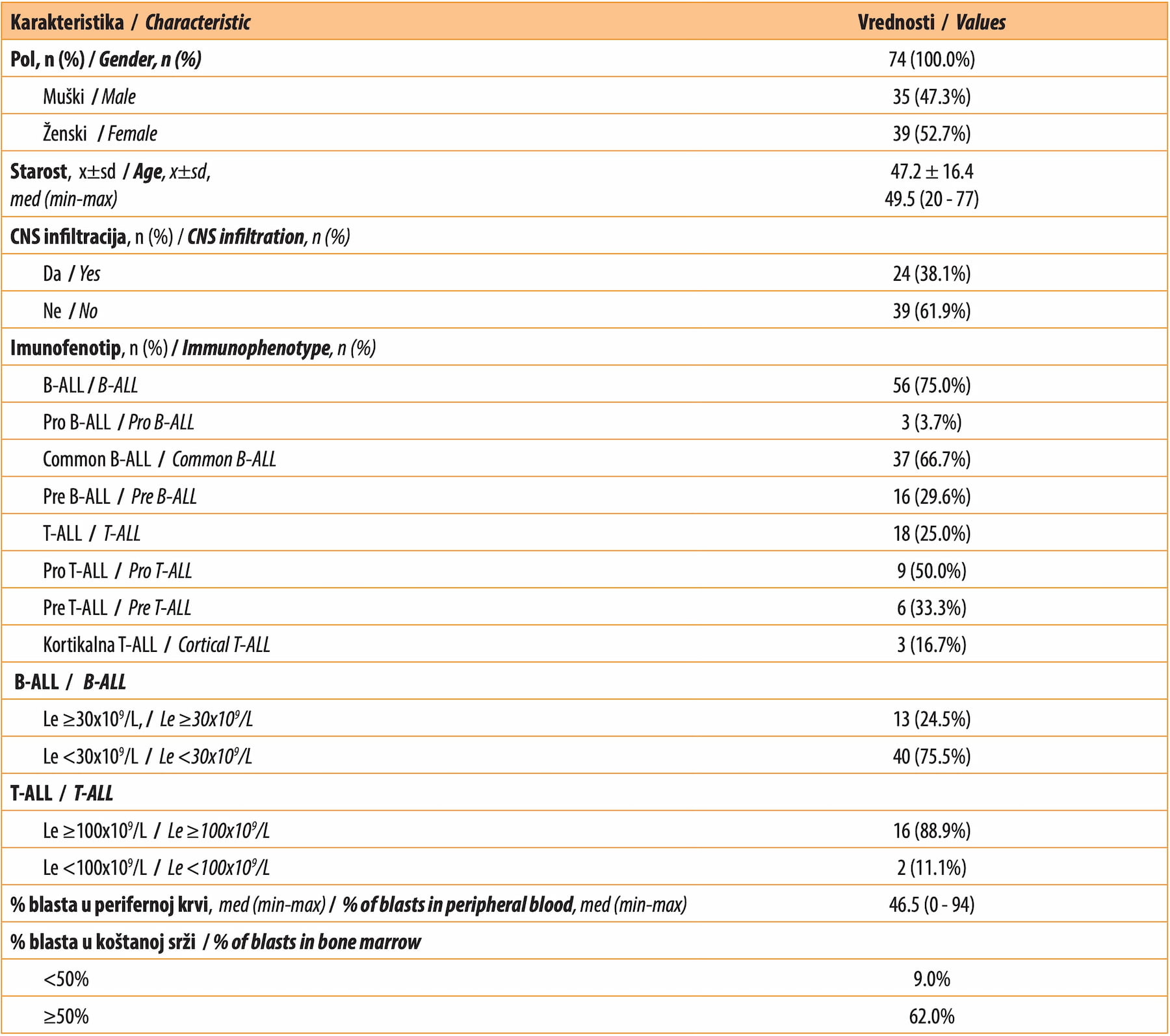
The study included 74 patients with ALL, of whom 75% suffered from B-cell ALL, while 25.0% were affected with T-cell ALL. The clinical and laboratory characteristics of the patients are presented in Table 1.
Most of the patients, 33 (47,1%) had a normal karyotype, while t(9;22) was present in 15 (23.5%) patients. Other chromosomal aberrations were present in 20 (29.4%) patients. PCR analysis determined the presence of BCR-ABL rearrangement in 22 (29.7%) patients; 50 (67.5%) patients did not have a positive PCR result for BCR-ABL, while there was no data for 2 (2.7%) patients.
The standard risk level was present in 20 (26.0%) patients; high risk was registered in 31 (45.2%) patients, while a very high risk was found in 22 (28.8%) patients.
A majority of the patients with B-cell ALL (75.5%) had a leukocyte count lower than 30x109 /L, while 24.5% had a leukocyte count above 30x109 /L. Amongst the patients with T-cell ALL, 88.9% had a leukocyte count above 100x109 /L, and only 11.1% patients had a leukocyte count lower than 100x109 /L.
The greatest number of patients with ALL, 63 (85.1%) of them, were treated with CT in keeping with the Hyper-CVAD protocol, while 11 (14.9%) patients were treated with the COP protocol. Hematopoietic stem cell transplantation was carried out in 17 (27.4%) patients, while 41 (70.7%) patients did not undergo transplantation.
In 54 (72.9%) patients, CR was achieved in 40 (71.4%) patients with B-cell ALL and 14 (77.7%) patients with T-cell ALL. Refractoriness to induction therapy was confirmed in 7 (9.4%) ALL patients, of whom 5 (8.9%) patients suffered from B-cell ALL, and 2 (11.1%) patients had T-ALL. Mortality in induction was registered in 13 (17.56%) patients, of whom 8 (14.2%) patients had B-cell ALL and 5 (27.7%) patients had T-cell ALL. Relapse occurred in 25 (46.3%) patients, i.e., 20 (47.5%) patients with B-cell ALL and 5 (33.3%) with T-cell ALL. Statistically significant difference between patients with B-cell ALL and those with T-cell ALL was not found for any of the analyzed parameters.
The median survival of ALL patients was 8 months, while five-year survival was registered in 23.2% subjects (Graph 1).
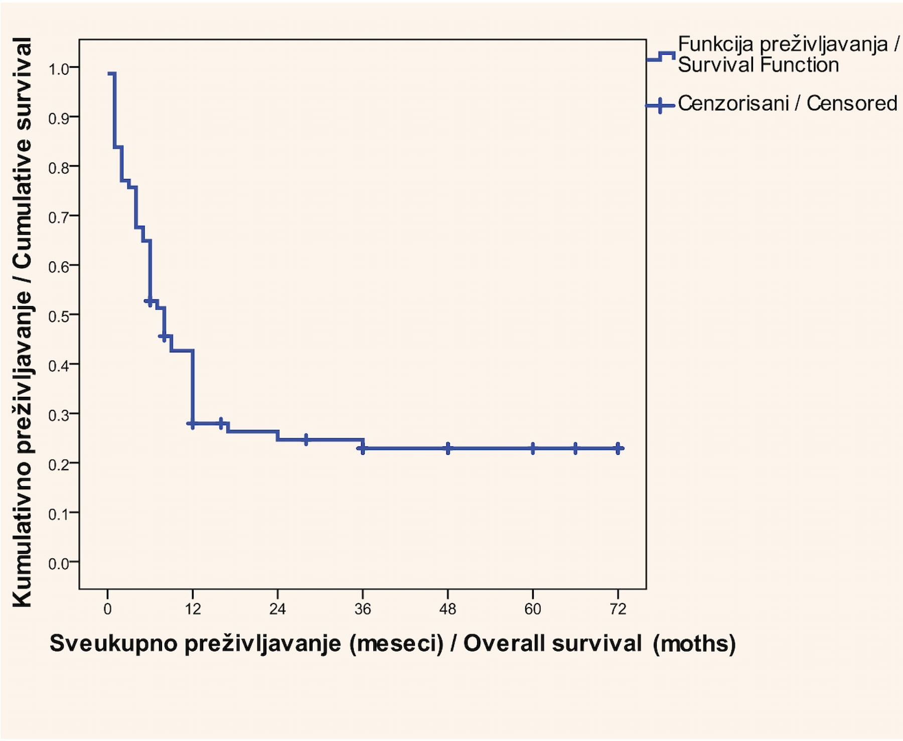
Graph 1. Overall survival of All patients by Kaplan-Meier method
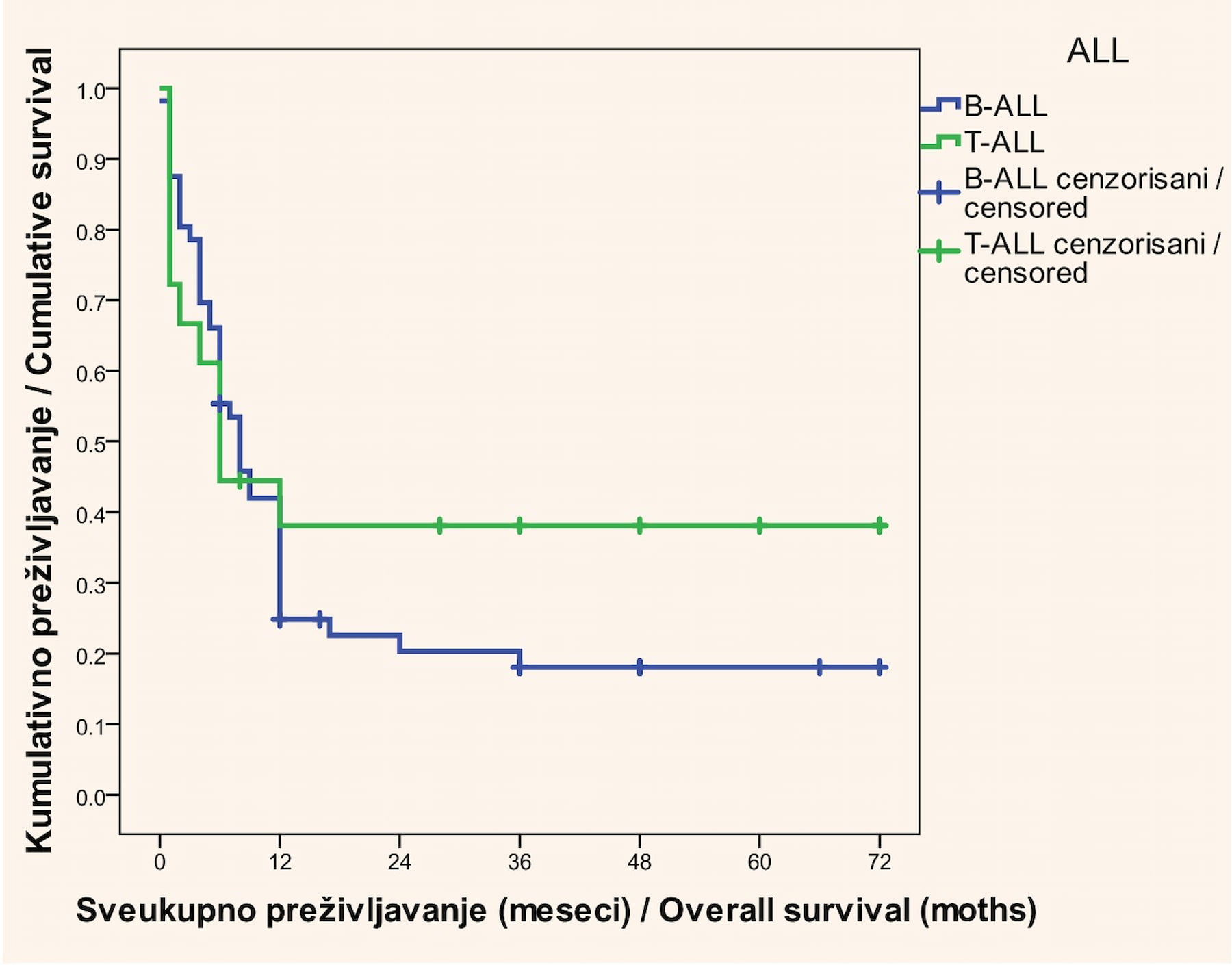
Median survival without signs of disease was 12 (1 - 72) months. The analysis of overall survival, according to the type of leukemia, showed that median survival in patients with B-cell ALL was 8 months, while it was 6 months in patients with T-cell ALL. Statistically significant difference in the survival time between B-cell and T-cell leukemia patients was not registered (p = 0.372) (Graph 2).
With the aid of the univariate Cox regression analysis, it was established that the following parameters had a statistically significant impact on poor overall survival: age ≥50 years (p < 0.001), application of the COP chemotherapy protocol (p < 0.001), non-transplant patients (p = 0.011), and CNS infiltration (p < 0.001) (Table 2).

Due to the unfavorable relationship between the outcome and the potential predictors, multivariate analysis was not performed.
DISCUSSION
Acute lymphoblastic leukemia is a malignant disease which occurs with a significantly poorer prognosis in adult age than in the pediatric population [2]. In addition to age, numerous factors, amongst which the most important are cytogenetic molecular changes, affect the outcome of the disease.
Our study included 74 patients, amongst whom the percentage of women (52.7%) was somewhat higher as compared to male subjects (47.3%). According to the data from literature, amongst ALL patients, male individuals are prevalent [14],[15].
In our test group, most of the patients had B-cell ALL; the common B-ALL immunological subtype - 37 (66.7%). The results of previous studies indicate that B-cell ALL is much more frequent than T-cell ALL and that it is present in 88.0% ALL patients, while T-cell ALL is present in 12.0% of ALL patients [16].
PCR analysis confirmed BCR-ABL positivity in 22 (29.7%) patients, of whom 4 (18.2%) had a normal karyotype, while in 18 (81.8%) patients t(9;22) was registered. The presence of the Philadelphia chromosome in ALL patients is considered to be a poor prognostic factor and is connected with shorter complete remission, grater refractoriness to the applied therapy, and poor overall survival. The results of other studies show that the Philadelphia chromosome occurs in 20% - 30% of the ALL adult patient population, while in the pediatric population, the frequency of its occurrence is lesser, amounting to 5.0% [17].
The leukocyte count is an important prognostic factor in acute lymphoblastic leukemia. In our study, 13 (24.5%) patients with B-cell ALL had a leukocyte count above 30x109 /L, while 16 (88.9%) patients with T-cell ALL had a leukocyte count above 100x109 /L. It is considered that a leukocyte count above 30x109 /L in B-cell ALL, as well as a leukocyte count above 100x109 /L in T-cell ALL, represents a poor prognostic factor. This is confirmed by a study by Rowe et al., showing that a leukocyte count above 30x109 /L presents an unfavorable prognostic factor [18].
The highest percentage of our patients, 63 (85.1%,), was treated with CT, according to the Hyper-CVAD protocol. Patients older than 65 years with an ECOG >2 and an HCT-CI >3 were treated with the COP protocol (11 patients – 14.9%). The results of previous studies show that a good response to the application of the Hyper-CVAD protocol was registered in 92.0% of the cases, with a 5-year survival rate of 32.0% [19].
CR was achieved in 54 (72.9%) patients, 40 (71.4%) patients with B-cell ALL and 14 (77.7%) patients with T-cell ALL. Mortality in induction was registered in 13 (17.5%) patients, among whom there were 8 (14.2%) patients with B-cell ALL and 5 (27.7%) patients with T-cell ALL. Relapse occurred in 25 (46.3%) patients, i.e., 20 (47.5%) patients with B-cell ALL and 5 (33.3%) patients with T-cell ALL. Refractoriness to induction therapy was confirmed in 7 (9.4%) ALL patients, of whom 5 (8.9%) had B-cell ALL, and 2 (11.1%) had T-cell ALL. The results from previous studies show that refractoriness is found in 10.0% of patients [17], while the early death rate ranges from 5.0% to 10.0%, while in the pediatric population it is significantly lower, amounting to 1.0% [20]. HSCT was caried out in 17 (27.4%) patients with a high and very high level of risk.
Bearing in mind that ALL is rarely registered in the adult population, there is little data indicating the survival rates of ALL patients. The study that was caried out demonstrated that the median overall survival of ALL subjects was 8 months, with somewhat longer survival in individuals with B-cell ALL, which did not prove to be as a statistically significant difference as compared to patients with T-cell ALL. Five-year survival in this study was 23.2%. In our study, median survival without signs of illness was 12 months. As compared to the results from our study, the rate of five-year survival was higher in American (35.5%) and German (43.4%) test groups [21].
Univariate analysis established that, in addition to age, the modality of applied therapy and CNS involvement were poor prognostic factors. These results have been confirmed by other studies demonstrating that older age is linked to a poorer prognosis, as well as that survival rates significantly decrease with increased age [21]. Patients aged between 15 and 24 years have a 10.0% higher rate of five-year survival as compared to patients aged between 25 and 39 years [5]. In a study by Lazarus et al., a statistically significant difference in overall survival was established in patients who had CNS involvement at diagnosis [22].
CONCLUSION
Our study has shown poor survival of ALL patients, which is significantly affected by age, the modality of administered therapy, and the involvement of the CNS. Introducing more precise molecular diagnostics, monitoring minimal residual disease, and the application of targeted therapy may contribute to a better survival of these patients, which has been confirmed by the latest research.
Informations

-
Keywords:
-
Received:
-
Revised:
-
Accepted:
-
Online first:
-
DOI:
-
Cite this article:

Ivana Damnjanović
General Hospital Valjevo, Valjevo, Serbia
2 Milenka Stojkovića Street, 14 000Valjevo, Serbia
E-mail:
1. Terwilliger T, Abdul-Hay M. Acute lymphoblastic leukemia: a comprehensive review and 2017 update. Blood Cancer J. 2017 Jun 30;7(6):e577. doi: 10.1038/bcj.2017.53.[CROSSREF]
2. Hunger SP, Lu X, Devidas M, Camitta BM, Gaynon PS, Winick NJ, et al. Improved survival for children and adolescents with acute lymphoblastic leukemia between 1990 and 2005: a report from the children's oncology group. J Clin Oncol. 2012 May 10;30(14):1663-9. doi: 10.1200/JCO.2011.37.8018.[CROSSREF]
3. Arber DA, Orazi A, Hasserjian R, et al. The 2016 revision to the World Health Organization classification of myeloid neoplasms and acute leukemia. Blood. 2016;127(20):2391-2405. Blood. 2016 Jul 21;128(3):462-3. doi: 10.1182/blood-2016-06-721662.[CROSSREF]
4. Stelljes M, Marks DI. Acute Lymphoblastic Leukemia in Adults. In: Carreras E, Dufour C, Mohty M, Kröger N, editors. The EBMT Handbook. Munster, Germany;2019;531-8.
5. Rowe JM. Prognostic factors in adult acute lymphoblastic leukaemia. Br J Haematol. 2010 Aug;150(4):389-405. doi: 10.1111/j.1365-2141.2010.08246.x.[CROSSREF]
6. Chiaretti S, Zini G, Bassan R. Diagnosis and subclassification of acute lymphoblastic leukemia. Mediterr J Hematol Infect Dis. 2014 Nov 1;6(1):e2014073. doi: 10.4084/MJHID.2014.073.[CROSSREF]
7. Gökbuget N, Dombret H, Ribera JM, Fielding AK, Advani A, Bassan R, et al. International reference analysis of outcomes in adults with B-precursor Ph-negative relapsed/refractory acute lymphoblastic leukemia. Haematologica. 2016 Dec;101(12):1524-33. doi: 10.3324/haematol.2016.144311. Epub 2016 Sep 1.[CROSSREF]
8. Bassan R, Spinelli O, Oldani E, Intermesoli T, Tosi M, Peruta B, et al. Improved risk classification for risk-specific therapy based on the molecular study of minimal residual disease (MRD) in adult acute lymphoblastic leukemia (ALL). Blood. 2009 Apr 30;113(18):4153-62. doi: 10.1182/ blood-2008-11-185132.[CROSSREF]
9. Vardiman JW, Brunning RD, Arber DA, et al. Introduction and overview of the classification of the myeloid neoplasms. In: Swerdlow SH Campo E, Lee Harris N, Jaffe ES, et al. (editors). WHO classification of tumours of hematopoietic and lymphoid tissues. IARC Lyon 2008;18:127-9.
10. Peterson LC, Bloomfield CD, Niemeyer CM, Döhner HGL. Myeloid neoplasms with germline predisposition. In: Swerdlow SH, Campo E, Harris NL, et al., editors. WHO Classification of Tumours of Haematopoietic and Lymphoid Tissues (Revised 4th ed). Lyon, IARC. 2017.
11. Gokbuget N, Arnold R, Bohme A, et al. Improved outcome in high risk and very high risk ALL by risk adapted SCT and in standard risk ALL by intensive chemotherapy in 713 adult ALL patients treated according to the prospective GMALL study 07/2003. ASH Annual Meeting Abstracts. 2007;110:12.
12. Béné MC, Nebe T, Bettelheim P, Buldini B, Bumbea H, Kern W, et al. Immunophenotyping of acute leukemia and lymphoproliferative disorders: a consensus proposal of the European LeukemiaNet Work Package 10. Leukemia. 2011 Apr;25(4):567-74. doi: 10.1038/leu.2010.312.[CROSSREF]
13. International System for Human Cytogenetic Nomenclature (ISCN). Recommendations for the International Standing Committee on Human Cytogenetic Nomenclature. Memphis, Tennessee, USA 1994.
14. Salah R, Musa HH, Hamid GA. Epidemiological study of acute lymphoblastic leukemia in Yemen. Eur J Pharm Sci 2017;4:794-7.
15. Santoyo-Sánchez A, Ramos-Peñafiel CO, Saavedra-González A, González-Almanza L, Martínez-Tovar A, Olarte-Carrillo I, et al. Frecuencias de edad y género de pacientes con leucemia en dos centros de referencia del Valle de México [The age and sex frequencies of patients with leukemia seen in two reference centers in the metropolitan area of Mexico City]. Gac Med Mex. 2017 Jan-Feb;153(1):44-8. Spanish.
16. Wartenberg D, Groves FD, Adelman AS. Acute lymphoblastic leukemia: epidemiology and etiology. In: Estey EH, Faderl S, Kantarjian H, editors. Acute Leukemias. 1. Berlin, Germany: Springer; 2008;77-93
17. Moorman AV, Harrison CJ, Buck GA, Richards SM, Secker-Walker LM, Martineau M, et al. Karyotype is an independent prognostic factor in adult acute lymphoblastic leukemia (ALL): analysis of cytogenetic data from patients treated on the Medical Research Council (MRC) UKALLXII/Eastern Cooperative Oncology Group (ECOG) 2993 trial. Blood. 2007 Apr 15;109(8):3189-97. doi: 10.1182/blood-2006-10-051912.[CROSSREF]
18. Rowe JM, Buck G, Burnett AK, Chopra R, Wiernik PH, Richards SM, et al. Induction therapy for adults with acute lymphoblastic leukemia: results of more than 1500 patients from the international ALL trial: MRC UKALL XII/ECOG E2993. Blood. 2005 Dec 1;106(12):3760-7. doi: 10.1182/blood-2005-04-1623.[CROSSREF]
19. Kantarjian HM, O’Brien S, Smith TL, Cortes J, Giles FJ, Beran M, et al. Results of treatment with hyper-CVAD, a dose-intensive regimen, in adult acute lymphocytic leukemia. J Clin Oncol. 2000 Feb;18(3):547-61. doi: 10.1200/JCO.2000.18.3.547.[CROSSREF]
20. Pui CH, Evans WE. Treatment of acute lymphoblastic leukemia. N Engl J Med. 2006 Jan 12;354(2):166-78. doi: 10.1056/NEJMra052603.[CROSSREF]
21. Pulte D, Jansen L, Gondos A, Katalinic A, Barnes B, Ressing M, et al. Survival of adults with acute lymphoblastic leukemia in Germany and the United States. PLoS One. 2014 Jan 27;9(1):e85554. doi: 10.1371/journal.pone.0085554.[CROSSREF]
22. Lazarus HM, Richards SM, Chopra R, Litzow MR, Burnett AK, Wiernik PH, et al. Central nervous system involvement in adult acute lymphoblastic leukemia at diagnosis: results from the international ALL trial MRC UKALL XII/ECOG E2993. Blood. 2006 Jul 15;108(2):465-72. doi: 10.1182/blood-2005-11-4666.[CROSSREF]