


Arthroscopically assisted osteosynthesis of tibial plateau fracture, Schatzker type III - Case report
- Institute for Orthopedic Surgery “Banjica”, Belgrade, Serbia
- Faculty of Medicine, University of Belgrade, Belgrade, Serbia
ABSTRACT
Introduction. Tibial plateau fractures are complex injuries that, in most cases, require surgical treatment in order to prevent far-reaching consequences for the functionality and quality of life of the patient. While numerous surgical techniques have been described, it is evident that the technique of arthroscopically assisted repositioning (reduction) and internal fixation has the potential to take the lead in the management of these types of injuries.
Case report. We present the case of a 36-year-old patient injured in a traffic accident with a fracture of the left tibial plateau, type III by Schatzker classification. After the diagnostic procedures and preparation, the patient was operated on using arthroscopically assisted repositioning (reduction) and internal fixation, with the filling of the bone defect with a combination of allogenic and synthetic bone graft. No other intra-articular injuries were diagnosed intraoperatively. The postoperative recovery went without complications, and after a period of 10 weeks of non-weight bearing and functional rehabilitation, the fracture healed completely, while the patient regained full range of motion in the knee.
Conclusion. Although this type of injury has traditionally been treated with open surgery, we believe that minimally invasive surgery is the future, when it comes to tibial plateau fractures Schatzker types I-III. There is still not enough data in literature regarding the long-term outcomes of this type of treatment, but the early postoperative results are extremely encouraging, since this surgical technique enables faster recovery and fewer postoperative complications.
INTRODUCTION
Fractures of the proximal tibia are complex injuries which involve damage to the osteal and soft tissue structures of the knee. They most commonly occur due to the impact of high intensity vertical force and/ or as the result of great valgus/varus stress in the knee joint. Untreated or improperly treated fractures lead to the development of secondary joint incongruency and degenerative disease [1],[2]. Despite a relatively low incidence of these injuries, their appropriate treatment is of great significance as the patients are, in most cases, members of the active work force [1],[3].
The main goal in the treatment of these injuries is establishing the best possible, hopefully anatomical, repositioning of the tibial joint surface and achieving ligamentous stability of the joint, while also achieving knee axis alignment and a full scope of movement in the joint [4]. As these are intra-articular fractures, such goals are attainable almost exclusively with the surgical method.
At present, there is a certain dissensus as to which surgical technique should be applied in the treatment of these fractures. The procedures most commonly applied are open reduction and internal fixation – ORIF, and arthroscopically assisted reduction and internal fixation - ARIF [1],[4],[5],[6],[7].
There is a number of classifications of these fractures, the two clinically most significant being the AO classification and the Schatzker classification [8],[9]. Simpler fracture types (Schatzker types I-III) can be treated both with the ORIF and with the ARIF surgical technique, while more severe fractures (Schatzker types IV-VI) are usually resolved with open surgery, although there are authors who feel that ARIF surgery can also be carried out in such cases [10].
The advantages of ARIF surgery are as follows: direct view of the joint surface during the entire procedure, possibility of arthroscopic treatment of the associated intraarticular osteal and soft tissue injuries, lesser trauma to the surrounding tissue, swifter postoperative recovery, and fewer postoperative complications [1],[10],[11],[12]. On the other hand, the advantages of open surgery lie in the possibility of treating all types of fractures, especially complex fractures or fractures in patients with osteoporosis, in whom the repositioning itself is challenging, while stabilization is possible with the application of buttress plates or locking plates [13],[14],[15]. Independently of the choice of surgical technique, poorly treated fractures have significant and long-term effects on the function and overall satisfaction of the patient, as well as on the increased risk of the development of secondary degenerative disease, which requires arthroplasty [16],[17].
The aim of this paper is to present the case of a female patient with a fracture of the left tibial plateau, type III by Schatzker classification, treated by the application of the method of arthroscopically assisted repositioning (reduction) and internal fixation, and to present the results of the early postoperative rehabilitation of this patient.
CASE REPORT
A 36-year-old patient was admitted to the ER with an injury to the left knee sustained in a car accident after direct impact of the lateral side of the left knee with the vehicle. At admission, she complained of diffuse pain in the region of the left knee, which was exacerbated by putting weight on it, and of restricted movement in the knee, caused by pain. On examination, a moderate swelling was noted in the anterolateral aspect of the knee and the lower leg, with the presence of a hematoma on the posterolateral side of the proximal end of the lower leg. On the lateral side of the lower leg there was an abrasion, 1 cm x 0.5 cm in size, which was not in communication with the deeper structures. Active and passive movement in the knee joint was limited by pain – flexion of around 20º, and extension 180º. Ligament stability tests were prevented by pain. Neurological and vascular findings of the left leg were normal, with palpable pulsation above the magistral blood vessels. Clinically, there were no signs of compartment syndrome of the lower leg. The patient had no other injuries.
After the examination, planar radiography of the left knee was performed in two directions -anteroposterior (AP) and left lateral (LL). A fracture of the lateral condyle of the tibia with a sinking of the broken fragment (type III fracture, by Schatzker classification), without signs of bone injury in the surrounding structures, was noted on the radiographic images. For the purpose of more precise evaluation and fracture classification, as a part of further diagnostics, computerized tomography (CT) was performed. An isolated fracture of the lateral tibial plateau, with central sinking, was noted on the CT images. The absence of other bone injury was confirmed (Figure 1).

Immediately after the diagnostics, an above the knee plaster-of-Paris splint (meniscus splint) was placed, and the patient was admitted to hospital for surgical treatment. For the purpose of assessing the condition of the soft tissue in the injured region and reducing the observed swelling, which could have jeopardized the planned therapeutic procedure, the patient was not operated on immediately after admission. The patient was advised to walk with the aid of crutches without putting any weight through the injured leg, until surgery. The following therapy was administered: prophylactic thromboembolic therapy – 3,800 IU of nadroparin calcium, administered subcutaneously, twice a day; analgesic therapy – 1,000 mg of paracetamol, four times a day in slow intravenous infusion, with additional administering of 2.5 g of metamizole sodium, in slow intravenous infusion, in periods of pain exacerbation.
On the fourth day after injury, once the swelling and the hematoma in the injured region had subsided, the operation was performed under general endotracheal anesthesia. Prophylactic antibiotic therapy was administered perioperatively in the form of slow intravenous infusion of a single dose of 1,000 mg of ceftriaxone. Just before the administering of anesthesia, stability of the knee was established in the sagittal and the frontal pane, with the application of clinical tests. The patient was placed to lie on her back, with the injured leg resting on the surgical leg holder at the level of the middle of the left lower leg, making it possible to perform full passive knee flexion. The right leg was positioned in abduction. A pneumatic tourniquet was positioned above the knee, and was, after extremity elevation and compressive exsanguination, pumped up to the internal pressure of 220 mmHg.
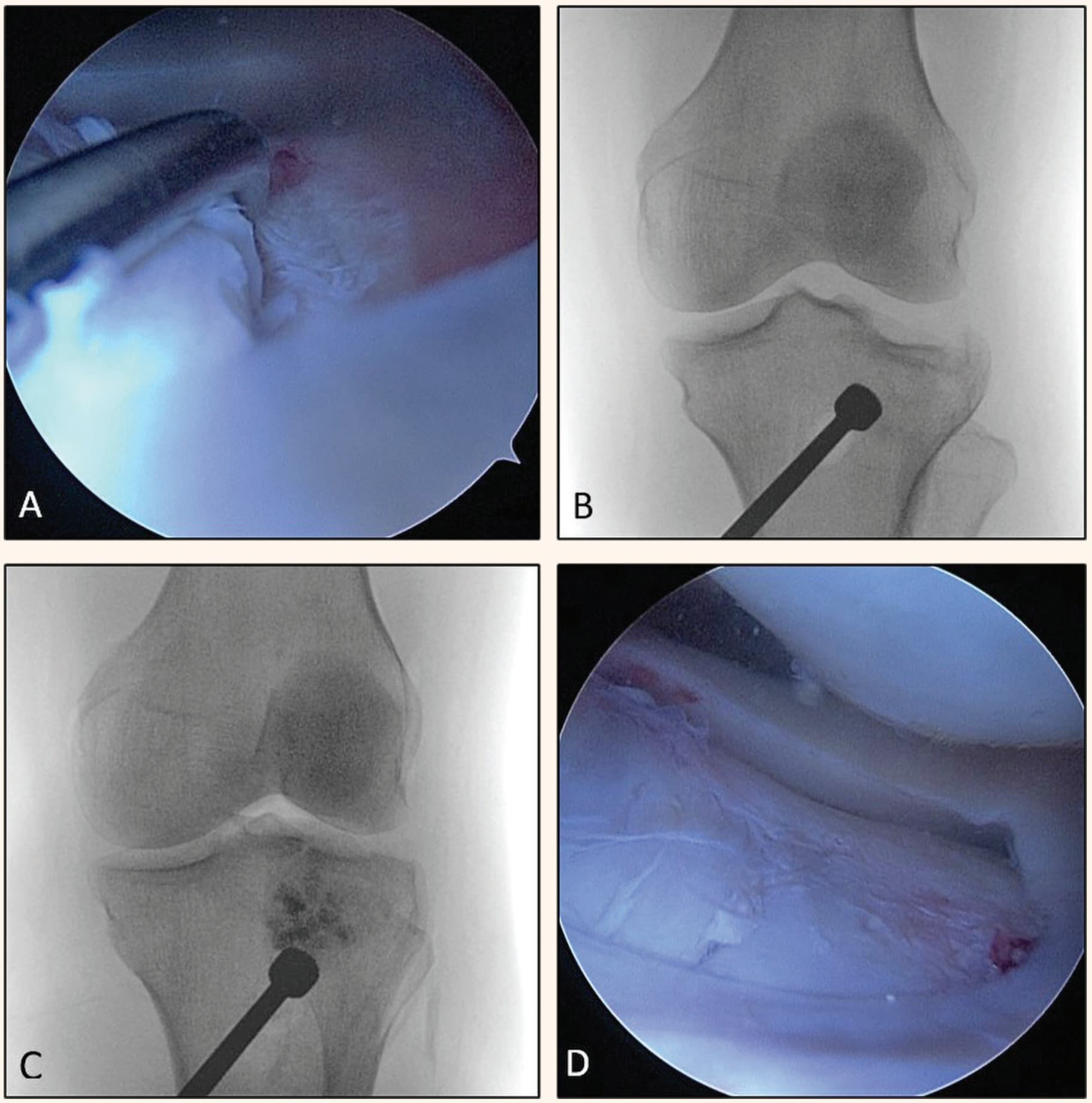
After the operative field was prepared, the knee joint was accessed via the two standard arthroscopic portals: the anterolateral (AL) and the anteromedial (AM). Hemarthrosis was determined arthroscopically, which is why the joint was rinsed with saline solution under pressure. An impression fracture of the lateral tibial plateau was detected (Figure 2.A). Arthroscopically, no pathological changes were found on the remaining osteal and soft tissue structures.
Entry was made via the AM portal with the standard tibial guide for the reconstruction of the anterior cruciate ligament (ACL), and its tip, controlled by an optical instrument was placed above the identified depression of the lateral condyle. At the site of the inferior end of the guide, at the level of the medial tibial plateau, a vertical incision was made into the skin and subcutaneous tissue, around 2 cm in length, and the tip of the guide was leaned against the cortex. With the control of an optical instrument and fluoroscopy a guide-needle was introduced via the guide to the middle of the depression of the lateral plateau, taking care not to rupture the joint cartilage. After the removal of the tibial guide, a drill, 10 mm in diameter, used to penetrate the cortex and enter the metaphyseal region, was positioned via the guide-needle. Then, the depression of the lateral plateau was repositioned with the aid of a blunt instrument and a hammer, under direct control of fluoroscopy and an optical instrument (Figure 2.B).
A mixture of bone graft from the bone bank and synthetic bone graft substitute was implanted into the metaphyseal region via the canal created in the bone, in order to fill the subchondral bone defect. The remainder of the canal was filled with the mixture up to the level of the cortex (Figure 2.C and 2.D).
With the use of fluoroscopic control, two parallel needles were introduced subcutaneously from the lateral side of the tibial plateau, directed medially, subchondrally and parallel to the joint surface. Through these needles, two cannulated partially threaded screws, 6.7 mm x 70 mm, were introduced, with foundations. After that, a follow-up radiography was carried out in order to check the position of the rafting screw. After the fixation of the fracture, stability was checked through knee joint movement (Figure 3).

The remaining tissue debris was arthroscopically removed from the joint cavity, and, after a test of hemostasis, an intraarticular drain was placed, after which a suture with individual stiches was made. Then, a cocktail with analgesic and hemostatic effects (8 ml of 0.5% bupivacaine injection solution, 1 ml of 1% adrenalin solution and 500 mg of tranexamic acid solution) was injected into the joint cavity. After the wounds were dressed with a sterile bandage, an elastic bandage was applied in order to prevent venostasis, upon which an above the knee plaster-of-Paris splint was placed, and the surgically treated leg was elevated while the patient was in bed.
Early postoperative recovery was normal, without local or systemic complications. After initial observation in the intensive care unit, the patient was transferred to the hospital ward and, on the same day, she was verticalized and enabled to walk with the help of two crutches, without putting weight through the surgically treated leg.
On the first postoperative day the drain was removed, and the patient was started on a regimen of passive exercises for the purpose of increasing the scope of movement in the knee. On the second postoperative day, the patient was discharged and was advised to walk using both crutches, without putting weight through the surgically treated leg, for the following 10 weeks. She was also advised to continue with the passive exercises for increasing the scope of movement in the surgically treated knee. The plaster-of-Paris splint was replaced with a functional knee orthosis in unlocked knee flexion mode. The patient was advised to lie down, elevate the leg and massage the knee with cold compresses after exercises, or after a longer period of time spent standing up or walking, in order to reduce swelling.
During postoperative hospital treatment, prophylactic thromboembolic therapy was continued – 3,800 IU of nadroparin calcium, administered subcutaneously, twice a day, as well as analgesic therapy – 1,000 mg of paracetamol, four times a day, 30 mg of ketorolac, three times a day, with additional administering of 2.5 g of metamizole sodium, in periods of pain exacerbation. At discharge, thromboembolic therapy was prescribed – rivaroxaban tablets, 15 mg, once a day, until the end of the 21st day after surgery, as well as analgesic therapy – etoricoxib, 90 mg, once a day in the first three days, and after that only in case of intense pain. Local application of heparin sodium, 1,000 IU/g in the form of a gel was advised, three times a day, as well as massage of the knee with ice, three to four times a day, at regular intervals, until the end of the 30th postoperative day.
The first follow-up examination was performed 12 days after the procedure, when the sutures from the patient’s surgical cuts were removed. During her recovery at home, the patient adhered to the instructions given to her at hospital discharge. The scars from the operation showed no sign of inflammation or infection and healed per primam intentionem. A mild diffuse swelling of the knee was registered. During the examination, passive flexion of the surgically repaired knee of 90º was performed, while extension was lacking by 5º. The neurological and vascular findings of the left leg were normal. Follow-up radiography confirmed a satisfactory position of the broken fragments and the implanted osteosynthetic material. The patient was advised to continue walking with two crutches without putting any weight through the injured leg, as well as to intensify the rehabilitation she had already started so as to increase the scope of movement in the knee and to strengthen the upper leg musculature.
The second follow-up examination was carried out six weeks after the operation. Through clinical examination, active flexion of 140º and extension up to -5º were noted. Follow-up radiography showed healing of the fracture in unchanged position.
The third follow-up examination was performed 10 weeks after surgery. Clinically, full painless movements were noted, while radiographically, healing of the fracture was confirmed in unchanged position (Figure 4). The patient was advised to discontinue wearing the functional orthosis and to start walking with the aid of crutches with partial weight-bearing through the surgically treated leg during the following two weeks, and after that gradual rejection of the orthopedic aids and the establishing of a normal walking regimen with full weight-bearing. The next follow-up was planned for 16 weeks after surgery, when gradual return to sports activities of low intensity might be planned.

DISCUSSION
Fractures of the tibial plateau are a therapeutic challenge, even for an experienced surgeon. This is due to the complexity of these fractures, the wide array of associated soft-tissue injuries, the demanding surgical technique, as well as invalidity occurring as the consequence of untreated or inadequately treated fractures, which is sometimes impossible to avoid even after very precisely executed surgical procedures. As in most intraarticular fractures, the best results are expected after surgical treatment, however, in a certain number of patients, the method of choice is nonsurgical treatment.
Nonsurgical treatment is applied in elderly patients with markedly osteoporotic bones, with low functional requirements; in patients with numerous comorbidities, which render surgical treatment unacceptably risky, i.e., life-threatening; in non-displaced or minimally displaced fractures; as well as in the case of a lesser depression of the lateral tibial plateau, without axial deformity [14],[18]. In such patients, fractures are treated by immobilizing the leg with an above-the-knee plaster-of-Paris splint, or, if possible, with a hinged knee brace with longitudinal side reinforcements, which allows movement in the knee, as well as with thromboembolic and analgesic therapy and physical therapy. Walking with crutches without putting any weight through the injured leg is recommended for a period of at least four to eight weeks, depending on the type of fracture [14].
An isolated depression of the lateral tibial plateau of 10 or more millimeters, which covers only a small surface of the joint cartilage, can be treated successfully non-surgically. However, if it is accompanied with tibial plateau separation, or if a significant portion of the joint surface is affected, it frequently causes secondary valgus axial deformity, which is why such injuries are treated surgically [14]. Numerous studies have reported good to excellent results in the nonsurgical treatment of patients with tibial plateau fractures [19],[20],[21],[22]. In their study, Waddell et al. report that a depression or separation of the lateral tibial plateau smaller than 10 mm can be successfully treated non-surgically, and Honkonen et al., in their study, report that a separation of 5 mm and a depression of the lateral tibial plateau of up to 3 mm are well tolerated in non-surgically treated patients, while fractures of the medial condyle should not be treated nonsurgically [23],[24].
Nevertheless, the pervasive view is that these intraarticular fractures should be treated surgically, whenever possible. The most common indications for surgical treatment are the following: displaced, unstable fractures with bone fragment depression of more than 5 mm and axial deformity greater than 10º in the frontal plane, as compared to the other, healthy leg; also, fractures with metaphyseal-diaphyseal propagation or unstable bicondylar fractures, compound fractures, fractures with accompanying or impending compartment syndrome, fractures with accompanying neurovascular injuries, and fractures of the proximal tibia with accompanying fractures of the distal end of the femur (floating knee) [25]. Hung et al. have reported that they took, as indications for surgical treatment, varus ligament instabilities in full extension – in fractures of the medial tibial plateau, varus and valgus ligament instabilities greater than 10º - in lateral tibial plateau fractures, as well as depression of the tibial joint surface greater than 3 mm and/or tibial condyle separation greater than 5 mm, measured radiographically, prior to surgery [1].
The exact size of the joint surface depression is often difficult to measure on AP and LL X-ray images. Martin et al. state that, when different observers independently measure radiographic discrepancies, their measurements differ by 12 or more millimeters [26]. For this reason, some authors believe that the number of millimeters is a simplified and not always reliable method for determining surgical indications [14]. The size of the tibial joint surface depression can be measured precisely and with great certainty on CT scans. In this way, fractures are classified more precisely, and the choice of surgical technique is more reliable [15],[27]. Chan et al. have demonstrated significant changes in the preoperative plan upon introducing CT diagnostics to follow standard radiographic imaging of the knee [28].
Classical, open surgery is most often performed through the anterolateral access, with the need for submeniscal arthrotomy, for the purpose of a better view of the joint surface and better control of fracture reduction [29]. A large incision into the soft tissue and arthrotomy, which may be accompanied by a transversal section of the meniscus, may postoperatively cause numerous complications, including the following: disruption in proprioception, pain which upsets normal joint function, reduction of the scope of movement, knee stiffness, hematoma in the area of the surgical wound, postoperative wound discharge, prolonged wound healing, necrosis of the wound margins, wound dehiscence, and infection [1],[6],[11],[30]. In their studies, Savoie and Lachiewicz have reported good to excellent results with lateral plateau fractures treated with the ORIF technique [31],[32].
Since Caspari and Jennings first published their studies on series of patients treated with arthroscopically assisted osteosynthesis, in 1985, this surgical technique has become an increasingly accepted method in treating lateral tibial plateau fractures [33],[34].
Arthroscopically assisted surgery enables a better view of the joint surfaces, direct assessment and, at the same time, appropriate treatment of certain intraarticular and soft-tissue injuries, which, according to certain studies, occur in 30% to 71% of patients with tibial plateau fractures [11]. In addition, like any endoscopic procedure, this one also, when compared to classical, open surgery, has advantages, such as: a lesser number of postoperative complications, smaller incisions and scars, less blood loss, reduced postoperative swelling and hematoma, possibility of removing hematoma and tissue debris from the joint, reduced soft tissue trauma, and thereby, swifter and easier recovery [7],[29]. According to some studies, in tibial plateau fractures, intramedullary vascularization is jeopardized, while periosteal vascularization is mostly preserved [35]. Arthroscopically assisted percutaneous fracture fixation protects the periosteal vascularization, thereby reducing the risk of the fracture failing to heal, the risk of osteonecrosis and soft-tissue necrosis, as well as the risk of infection [36].
Fowble et al. have reported far better results in patients treated with ARIF methods as compared to those treated with ORIF [37]. Kiefer, Van Glabbeek, and Ohdera have shown good to excellent results with patients treated with arthroscopically assisted surgery [38],[39],[40]. With ARIF and ORIF techniques, good clinical and radiographic results have been achieved, without statistically significant differences in complication rates, while patients treated with arthroscopically assisted surgery were hospitalized for a shorter period of time, and achieved full weight bearing earlier [3],[11]. Certain authors feel that arthroscopic surgery is a very demanding procedure, in the technical sense, and that it should be carried out exclusively by an experienced surgeon and applied only in fractures of the lateral condyle of the tibia (Schatzker types I-III) [41].
Despite recommendations by certain studies to be aware of the fact that the use of a pump may lead to the development of the compartment syndrome [3],[11],[42], we decided to use that particular method of rinsing the knee joint, encouraged by the studies stating that this complication is rarer in fractures of the lateral tibial plateau, since, in this way, tissue debris is better removed from the joint fissure [43]. After rinsing the joint, we noted an isolated impression fracture of the lateral tibial plateau. There were no accompanying injuries of the bone and soft-tissue structures, as opposed to the study by Verona et al., where soft-tissue structures occurred in 52.5% of the patients, and a study by Kayali et al., where such injuries were present in 67% of patients [7],[11]. In their paper, Wang et al. have stated that fractures of the lateral tibial plateau were accompanied by some form of injury to the anterior cruciate ligament in around 80% of cases, out of which 48% had a complete lesion, while 48% of the subjects sustained injury to the lateral meniscus [44]. We filled the bone defect with a combination of allograft (graft from the bone bank) and synthetic bone graft substitute, while the fracture was stabilized with cannulated screws, positioned parallel to each other, subchondrally, by means of the rafting technique, as described by numerous authors [1],[7],[11],[14],[15],[36].
Immediately after the operation, the leg was immobilized and elevated, with the application of cold compresses on the injured knee, in keeping with the recommendations by Hung et al. [1]. Patients used immobilization from two days to twelve weeks, but most commonly from three to six weeks, which varies from study to study [45]. Due to insufficient experience by the surgeon in the use of this particular type of surgical technique for treating lateral tibial plateau injuries, in the case of our patient, we immobilized the knee with a functional knee orthosis which allows for full movement in the knee joint, for a period of 10 weeks. Passive increase of the scope of movement in the surgically treated knee and active isometric exercises of the upper leg muscles began on the first postoperative day, upon drain removal, in keeping with a study by Boldin et al. [46]. Active exercises for the surgically treated knee began after the removal of sutures.
According to the review by Arnold et al., in as many as 79% of the cases, the postoperative time of non-weightbearing walking was shorter than 10 weeks, while partial weightbearing was introduced, in 46% of the cases, between the fourth and the sixth postoperative week [45]. As there are no clear recommendations on introducing increasing levels of weight bearing in patients treated with the ARIF surgical technique, we decided on the more conservative approach, recommending our patient to walk with crutches without putting weight through the injured leg, for a period of 10 weeks, which is in keeping with the current AO recommendations and other larger studies [1],[45],[47]. Ten weeks after the operation, at the latest check-up, full scope of movement in the knee was achieved.
In the presented patient there were neither any iatrogenic injuries nor were there any intraoperative or early postoperative complications. We are aware that the follow-up period is short, and that rehabilitation is not yet over, however a good early functional result and the radiographic findings are encouraging as to the further application of this surgical procedure, motivating us to try and perfect it further and improve the surgical technique, as well as postoperative recovery. We will definitely continue to monitor the patient, both clinically and radiographically. Analysis and monitoring of a larger number of patients over a longer period of time would provide more relevant data, which would indicate whether arthroscopically assisted osteosynthesis of lateral tibial condyle fracture is an appropriate and cost-effective method.
Informations

-
Keywords:
-
Received:
-
Revised:
-
Accepted:
-
Online first:
-
DOI:
-
Cite this article:

Nemanja Jovanović
Institute for Orthopedic Surgery “Banjica”
28 Mihaila Avramovica Street, 11040 Belgrade, Serbia
E-mail:
1. Hung SS, Chao EK, Chan YS, Yuan LJ, Chung PC, Chen CY, et al. Arthroscopically assisted osteosynthesis for tibial plateau fractures. J Trauma. 2003 Feb; 54(2):356-63.[CROSSREF]
2. Benea H, Tomoaia G, Martin A, Bardas C. Arthroscopic management of proximal tibial fractures: technical note and case series presentation. Clujul Med. 2015; 88(2):233-6.[CROSSREF]
3. Elabjer E, Benčić I, Ćuti T, Cerovečki T, Ćurić S, Vidović D. Tibial plateau fracture management: arthroscopically-assisted versus ORIF procedure - clinical and radiological comparison. Injury. 2017 Nov; 48 Suppl 5:S61-S64.[CROSSREF]
4. Hermanowicz K, Malinowski K, Góralczyk A, Guszczyn T, LaPrade RF, Sadlik B. All-Arthroscopic Treatment of Schatzker Type III Lateral Tibial Plateau Fracture Without Fluoroscopy. Arthrosc Tech. 2019 May 17; 8(6):e567-e574.[CROSSREF]
5. Mthethwa J, Chikate A. A review of the management of tibial plateau fractures. Musculoskelet Surg. 2018 Aug; 102(2):119-27.[CROSSREF]
6. Le Baron M, Cermolacce M, Flecher X, Guillotin C, Bauer T, Ehlinger M. Tibial plateau fracture management: ARIF versus ORIF - clinical and radiological comparison. Orthop Traumatol Surg Res. 2019 Feb; 105(1):101-6.[CROSSREF]
7. Kayali C, Oztürk H, Altay T, Reisoglu A, Agus H. Arthroscopically assisted percutaneous osteosynthesis of lateral tibial plateau fractures. Can J Surg. 2008 Oct; 51(5):378-82.[Google Scholar]
8. Meinberg EG, Agel J, Roberts CS, Karam MD, Kellam JF. Fracture and Dislocation Classification Compendium-2018. J Orthop Trauma. 2018 Jan; 32 Suppl 1:S1-S170.[CROSSREF]
9. Schatzker J, McBroom R, Bruce D. The tibial plateau fracture. The Toronto experience 1968--1975. Clin Orthop Relat Res. 1979 Jan-Feb; (138):94-104.
10. Dall’oca C, Maluta T, Lavini F, Bondi M, Micheloni GM, Bartolozzi P. Tibial plateau fractures: compared outcomes between ARIF and ORIF. Strategies Trauma Limb Reconstr. 2012 Nov; 7(3):163-75.[CROSSREF]
11. Verona M, Marongiu G, Cardoni G, Piras N, Frigau L, Capone A. Arthroscopically assisted reduction and internal fixation (ARIF) versus open reduction and internal fixation (ORIF) for lateral tibial plateau fractures: A comparative retrospective study. J Orthop Surg Res. 2019 May 24; 14(1):155.[CROSSREF]
12. Robertson GAJ, Wong SJ, Wood AM. Return to sport following tibial plateau fractures: A systematic review. World J Orthop. 2017 Jul 18; 8(7):574-87.[CROSSREF]
13. Prat-Fabregat S, Camacho-Carrasco P. Treatment strategy for tibial plateau fractures: an update. EFORT Open Rev. 2017 Mar 13; 1(5):225-32.[CROSSREF]
14. Marsh JL, Karam MD. Tibial Plateau Fractures. In: Court-Brown CM, Heckman JD, McQueen MM, Ricci W, Tornetta P, McKee M, editors. Rockwood and Green’s fractures in adults. 8th ed. Philadelphia: Wolters Kluwer Health; 2015; p.2303-67.
15. Rudloff MI. Fractures of the Lower Extremity. In: Azar FM, Beaty JH, Canale ST, editors. Campbell’s Operative Orthopaedics. 13th ed. Philadelphia: Elsevier Inc; 2017; p.2712-816.
16. Singleton N, Sahakian V, Muir D. Outcome After Tibial Plateau Fracture: How Important Is Restoration of Articular Congruity? J Orthop Trauma. 2017 Mar; 31(3):158-63.[CROSSREF]
17. Wasserstein D, Henry P, Paterson JM, Kreder HJ, Jenkinson R. Risk of total knee arthroplasty after operatively treated tibial plateau fracture: A matched-population-based cohort study. J Bone Joint Surg Am. 2014 Jan 15; 96(2):144-50.[CROSSREF]
18. Rozell JC, Vemulapalli KC, Gary JL, Donegan DJ. Tibial Plateau Fractures in Elderly Patients. Geriatr Orthop Surg Rehabil. 2016 Sep; 7(3):126-34.[CROSSREF]
19. Drennan DB, Locher FG, Maylahn DJ. Fractures of the tibial plateau. Treatment by closed reduction and spica cast. J Bone Joint Surg Am. 1979 Oct; 61(7):989-95.[CROSSREF]
20. Lansinger O, Bergman B, Körner L, Andersson GB. Tibial condylar fractures. A twenty-year follow-up. J Bone Joint Surg Am. 1986 Jan; 68(1):13-9.[CROSSREF]
21. Scotland T, Wardlaw D. The use of cast-bracing as treatment for fractures of the tibial plateau. J Bone Joint Surg Br. 1981; 63B(4):575-8.[CROSSREF]
22. Duwelius PJ, Connolly JF. Closed reduction of tibial plateau fractures. A comparison of functional and roentgenographic end results. Clin Orthop Relat Res. 1988 May; (230):116-26.[CROSSREF]
23. Waddell JP, Johnston DW, Neidre A. Fractures of the tibial plateau: a review of ninety-five patients and comparison of treatment methods. J Trauma. 1981 May; 21(5):376-81.[CROSSREF]
24. Honkonen SE. Indications for surgical treatment of tibial condyle fractures. Clin Orthop Relat Res. 1994 May; (302):199-205.[CROSSREF]
25. Rajasekaran S, Kamath V, Dheenadhaylan J. Tibial Plateau Fractures. In: Sivananthan S, Sherry S, Warnke P, Miller MD, editors. Mercer’s Textbook of Orthopaedics and Trauma. 10th ed. London: Hodder Education; 2012; p.372-6.[CROSSREF]
26. Martin J, Marsh JL, Nepola JV, Dirschl DR, Hurwitz S, DeCoster TA. Radiographic fracture assessments: which ones can we reliably make? J Orthop Trauma. 2000 Aug; 14(6):379-85.[CROSSREF]
27. Dodd A, Oddone Paolucci E, Korley R. The effect of three-dimensional computed tomography reconstructions on preoperative planning of tibial plateau fractures: a case-control series. BMC Musculoskelet Disord. 2015 Jun 13; 16:144.[CROSSREF]
28. Chan PS, Klimkiewicz JJ, Luchetti WT, Esterhai JL, Kneeland JB, Dalinka MK, et al. Impact of CT scan on treatment plan and fracture classification of tibial plateau fractures. J Orthop Trauma. 1997 Oct; 11(7):484-9.[CROSSREF]
29. Kandemir U, Maclean J. Surgical approaches for tibial plateau fractures. J Knee Surg. 2014 Feb; 27(1):21-9.[CROSSREF]
30. Chen XZ, Liu CG, Chen Y, Wang LQ, Zhu QZ, Lin P. Arthroscopy-assisted surgery for tibial plateau fractures. Arthroscopy. 2015 Jan; 31(1):143-53.[CROSSREF]
31. Savoie FH, Vander Griend RA, Ward EF, Hughes JL. Tibial plateau fractures. A review of operative treatment using AO technique. Orthopedics. 1987 May; 10(5):745-50.[CROSSREF]
32. Lachiewicz PF, Funcik T. Factors influencing the results of open reduction and internal fixation of tibial plateau fractures. Clin Orthop Relat Res. 1990 Oct; (259):210-5.[CROSSREF]
33. Caspari RB, Hutton PM, Whipple TL, Meyers JF. The role of arthroscopy in the management of tibial plateau fractures. Arthroscopy. 1985; 1(2):76-82.[CROSSREF]
34. Jennings JE. Arthroscopic management of tibial plateau fractures. Arthroscopy. 1985; 1(3):160-8.[CROSSREF]
35. Hannouche D, Duparc F, Beaufils P. The arterial vascularization of the lateral tibial condyle: anatomy and surgical applications. Surg Radiol Anat. 2006 Mar; 28(1):38-45.[CROSSREF]
36. Burdin G. Arthroscopic management of tibial plateau fractures: surgical technique. Orthop Traumatol Surg Res. 2013 Feb; 99(1 Suppl):S208-18.[CROSSREF]
37. Fowble CD, Zimmer JW, Schepsis AA. The role of arthroscopy in the assessment and treatment of tibial plateau fractures. Arthroscopy. 1993; 9(5):584-90.[CROSSREF]
38. Ohdera T, Tokunaga M, Hiroshima S, Yoshimoto E, Tokunaga J, Kobayashi A. Arthroscopic management of tibial plateau fractures - comparison with open reduction method. Arch Orthop Trauma Surg. 2003 Nov; 123(9):489-93.[CROSSREF]
39. Kiefer H, Zivaljevic N, Imbriglia JE. Arthroscopic reduction and internal fixation (ARIF) of lateral tibial plateau fractures. Knee Surg Sports Traumatol Arthrosc. 2001 May; 9(3):167-72.[CROSSREF]
40. van Glabbeek F, van Riet R, Jansen N, D’Anvers J, Nuyts R. Arthroscopically assisted reduction and internal fixation of tibial plateau fractures: report of twenty cases. Acta Orthop Belg. 2002 Jun; 68(3):258-64.[Gooogle Scholar]
41. Tornetta P 3rd. Arthroscopic elevation with grafting. J Orthop Trauma. 2002 Jul; 16(6):444-6.[CROSSREF]
42. Belanger M, Fadale P. Compartment syndrome of the leg after arthroscopic examination of a tibial plateau fracture. Case report and review of the literature. Arthroscopy. 1997 Oct; 13(5):646-51.[CROSSREF]
43. Herbort M, Domnick C, Petersen W. Arthroscopic treatment of tibial plateaufractures. Oper Orthop Traumatol. 2014; 26:573–88.[CROSSREF]
44. Wang Y, Cao F, Liu M, Wang J, Jia S. Incidence of Soft-Tissue Injuries in Patients with Posterolateral Tibial Plateau Fractures: A Retrospective Review from 2009 to 2014. J Knee Surg. 2016 Aug; 29(6):451-7.[CROSSREF]
45. Arnold JB, Tu CG, Phan TM, Rickman M, Varghese VD, Thewlis D, et al. Characteristics of postoperative weight bearing and management protocols for tibial plateau fractures: Findings from a scoping review. Injury. 2017 Dec; 48(12):2634-42.[CROSSREF]
46. Boldin C, Fankhauser F, Hofer HP, Szyszkowitz R. Three-year results of proximal tibia fractures treated with the LISS. Clin Orthop Relat Res. 2006 Apr; 445:222-9.[CROSSREF]
47. Cong-Feng L. Tibia, proximal. In: Buckley RE, Moran CG, Apivatthakakul T, editors. AO Principles of Fracture Menagement, 3rd ed. New York: Thieme; 2017; p.877-98.
- Figure 1
- Figure 2. Arthroscopic image of the fracture (A), fluoroscopic image of the positioning of a blunt instrument into the canal in the bone (B), fluoroscopic image after the implantation of the combination of allogenic and synthetic bone graft substitute (C), arthroscopic image after repositioning (D)
- Figure 3. Postoperative follow-up radiographic images: AP (A) and LL (B)
- Figure 4. Follow-up radiography, 10 weeks after the operation: AP (A) and LL (B)