


The prevalence of metabolic disorders and hypertension in women during perimenopause and menopause
- University of Priština – Kosovska Mitrovica, Faculty of Medicine, Kosovska Mitrovica, Serbia
- University of Belgrade, Faculty of Medicine, Belgrade, Serbia
ABSTRACT
Introduction/Objective: Cardiometabolic diseases, including type 2 diabetes (T2D) and cardiovascular disease, and their associated factors, such as hypertension, dyslipidemia, insulin resistance, and obesity, are among the leading causes of morbidity and mortality. At a young age, the prevalence of cardiometabolic diseases is higher in men than in women, but this female advantage gradually disappears with aging, particularly after menopause, when cardiometabolic risk factors accumulate. Our research aimed to highlight the prevalence of metabolic disorders and their association with hypertension in women during perimenopause and menopause.
Method: The study included 200 middle-aged women: 131 (65.5%) perimenopausal women who had experienced typical perimenopausal signs for more than 6 months, and 69 (34.5%) menopausal women with absent menstrual cycles for more than 12 months, aged 50-59 years. The patients visited their physician for a routine preventive examination in December 2024.
Results: Hypertension was statistically significantly more common in women in menopause (84.1%) compared to premenopausal women (p < 0.001). Women in menopause had significantly higher rates of metabolic disorders, increased body mass and obesity, with 66.7% of the participants affected, as well as a larger waist circumference in 63.8% of the participants (p = 0.003). Menopausal women had statistically significantly higher levels of triglycerides, LDL cholesterol, and total cholesterol than premenopausal women (p = 0.002). Among women going through menopause, diabetes mellitus was statistically substantially more prevalent, affecting 27% of them (p = 0.002). Premenopausal women had a statistically significantly greater prevalence of hypothyroidism (14.7%) than did menopausal women (4.3%) (p = 0.027). These two groups of participants did not differ significantly in terms of sedentary habits, smoking, or family history of hypertension.
Conclusion: Type 2 diabetes, obesity, dyslipidaemia, and hypertension were all substantially more common in menopausal women. Preventive screenings are therefore crucial for women going through menopause and perimenopause to promptly detect and treat cardiometabolic disorders and prevent more serious cardiovascular diseases.
INTRODUCTION
Cardiometabolic diseases, including type 2 diabetes mellitus and hypertension, are among the leading causes of morbidity and mortality in Serbia. At a younger age, the prevalence of cardiometabolic diseases is higher in men than in women; however, this advantage gradually diminishes with aging, particularly during menopause, when women are at increased cardiovascular risk. Although reproductive aging is characteristic of both sexes, the stages of reproductive aging are better described in women. Estrogens are the prototype female hormones and exhibit full activity in the years preceding menopause. Menopause is a stage of life characterized not only by a decline in estrogen levels and a relative excess of androgens, but also by numerous psychophysiological changes resulting from hormonal imbalance [1].
It is expected that by 2025, there will be more than 1 billion women in menopause worldwide [2]. Women more often face inadequate diagnosis and treatment of cardiovascular diseases compared to men. Even when traditional cardiovascular risk factors are present in women, physicians do not attribute adequate importance to them, which leads to poorer disease outcomes. Hypertension is more common among women and carries a twofold higher risk of mortality compared to men [3]. Women with diabetes have a higher risk of ischemic heart disease, by 3 to 7 times, compared to men, in whom the risk is only 2 to 3 times higher in the presence of type 2 diabetes mellitus [4]. Type 2 diabetes mellitus has been confirmed in one out of nine women. Although there appears to be no correlation between the onset of menopause and higher glucose levels, an increased risk of metabolic syndrome emerges after the transition to menopause [5]. During menopause, additional factors significantly increase cardiovascular risk. A recent meta-analysis showed that smoking increases the relative risk of major cardiovascular events by 25% in women compared to men [6].
Sex-specific risk factors and their association with cardiovascular disease (CVD) risk are increasingly being studied and recognized, emphasizing the importance of a detailed reproductive and obstetric history for cardiovascular risk stratification. Sex-specific risk factors, including hypertensive disorders in pregnancy, gestational diabetes, preterm delivery and placental abruption, early menarche, early menopause and vasomotor symptoms in menopause, polycystic ovary syndrome, infertility and infertility treatment, autoimmune diseases, and depression, are associated with increased cardiovascular risk [7],[8].
Hypertension is a highly prevalent and powerful cardiovascular risk factor in women, associated with myocardial infarction, heart failure, stroke, atrial fibrillation, peripheral arterial disease, and chronic kidney disease. Systolic blood pressure begins to rise around the time of the menopausal transition and becomes more frequently elevated after the age of 65 in women compared to men. Young women with hypertension are at greater risk of target organ damage compared to men of the same age. The decline in estrogen levels leads to autonomic dysfunction, generalized endothelial dysfunction, and increased activity of the renin–angiotensin–aldosterone system, thereby contributing to elevated blood pressure [9],[10].
Dyslipidemia is a well-known risk factor for CVD, with a direct relationship between LDL cholesterol and atherosclerotic cardiovascular disease. In women, hyperlipidemia is more common during menopause. Thus, women in menopause more frequently have higher levels of total cholesterol, triglycerides, and LDL cholesterol, as well as lower levels of HDL cholesterol, placing them at higher risk for CVD [11],[12]. High levels of non-HDL cholesterol and triglycerides are more important risk factors for CVD in women than in men, particularly in women with diabetes. Increased body mass index (BMI) is associated with increased cardiovascular risk in both sexes; however, there are sex differences in fat distribution, including visceral and subcutaneous fat. During menopause, women experience an increase in visceral fat compared to premenopausal women of similar age, which contributes to insulin resistance and inflammation [1],[11],[12],[13].
Our study aimed to assess the prevalence of non-modifiable and modifiable risk factors and their association with metabolic disorders and hypertension in premenopausal and menopausal women.
METHODOLOGY
A prospective study included 200 middle-aged women who attended a routine preventive medical examination during December 2024.
The study was a cross-sectional design and included 200 middle-aged women who presented for a routine preventive examination in December 2024. All participants provided written informed consent to participate in the study, and the study was conducted in accordance with ethical standards.
The patients were divided into two groups. There were 131 (65.5%) women in perimenopause who had typical signs of perimenopause for more than 6 months, and 69 (34.5%) women in menopause with absence of menstruation for more than 12 months, aged between 50 and 59 years. Based on the gynecological history, the patients were classified into these two groups. The diagnosis of arterial hypertension was established according to the European Society of Hypertension recommendations.
Sociodemographic data were obtained from all patients, including age, place of residence, marital status, education, and occupation. Anamnestic data were also collected (previous diseases, family history, smoking, alcohol consumption, and level of physical activity). Laboratory analyses included complete blood count and biochemical parameters. A clinical examination was performed, along with additional diagnostic procedures, including ECG, echocardiography, and abdominal ultrasound.
The study was conducted in accordance with current ethical standards and the principles of Good Clinical Practice, as outlined in the Declaration of Helsinki.
STATISTICAL ANALYSIS
Statistical analyses were performed using SPSS Statistics version 22 (SPSS Inc., Chicago, IL, USA). Continuous variables did not have a normal distribution and were therefore presented as median (Min–Max), and differences between groups were tested using the Mann– Whitney test. Categorical variables were presented as numbers (percentages), and differences in frequency were tested using the chi-square test.
The criterion for statistical significance was p < 0.05.
RESULTS
The study included 200 female participants. The participants were divided into two groups. The first group consisted of premenopausal women younger than 49 years and menopausal women aged 50 to 59 years (Table 1).
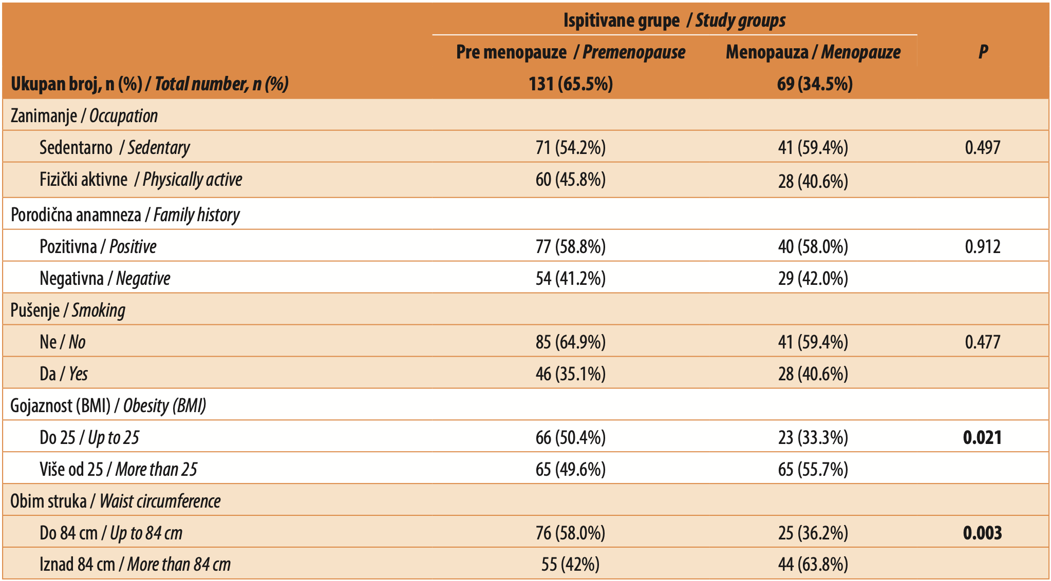
Sociodemographic characteristics were comparable between the groups. Occupation and level of physical activity were similar in both groups, as was cigarette smoking. There was a statistically significant difference in nutritional status and waist circumference. In menopausal women, obesity-related parameters were significantly more frequent, including BMI > 25 (p = 0.021) and waist circumference > 84 cm (p = 0.003) (Table 1).
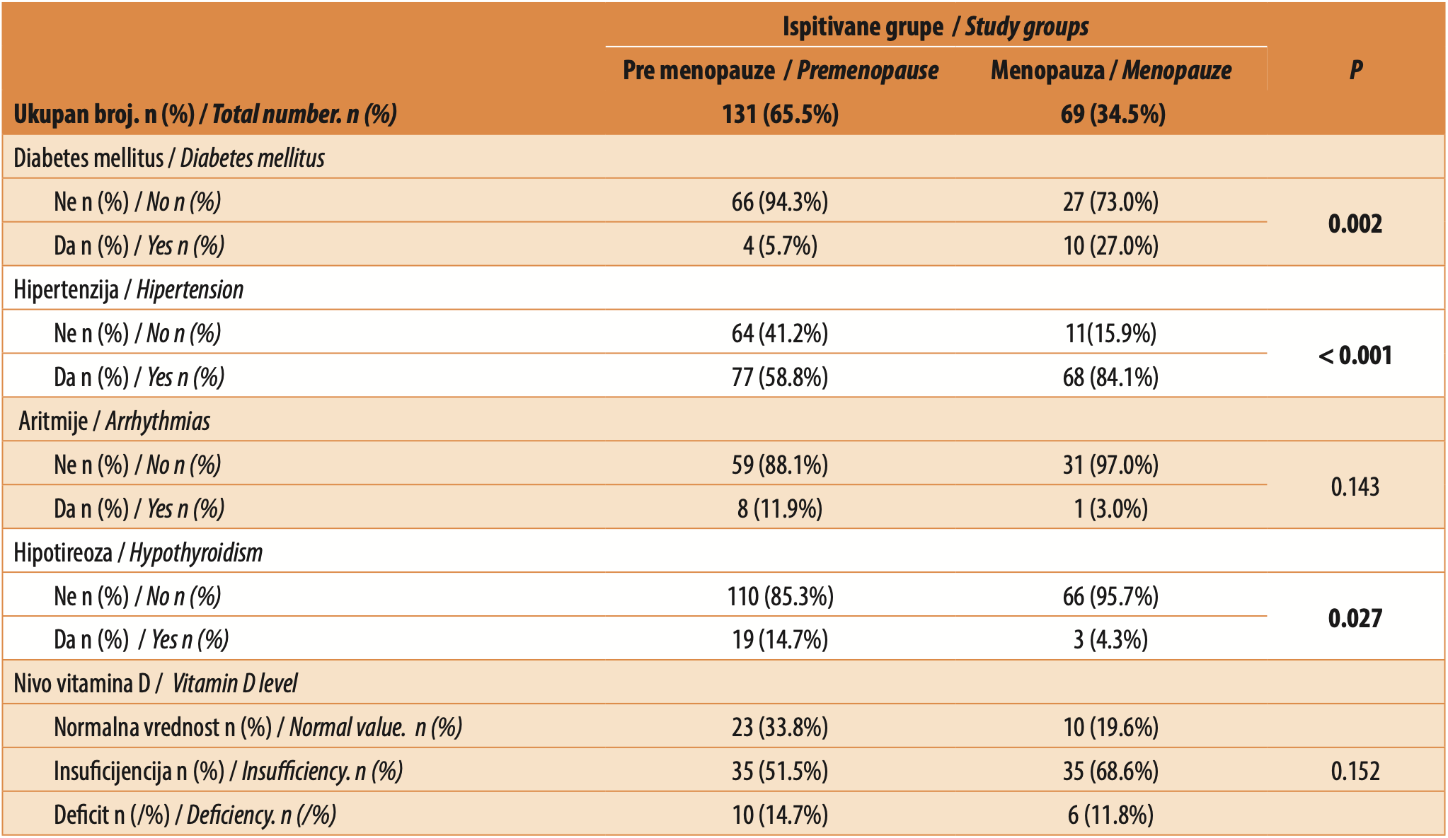
Women in menopause more frequently had hypertension (p < 0.001) and type 2 diabetes mellitus (p = 0.002). Arrhythmia was equally common in both groups, as was vitamin D deficiency. Hypothyroidism was significantly more frequent (p = 0.027) in the younger group of women in perimenopause, and all of them were receiving substitution therapy (Table 2).
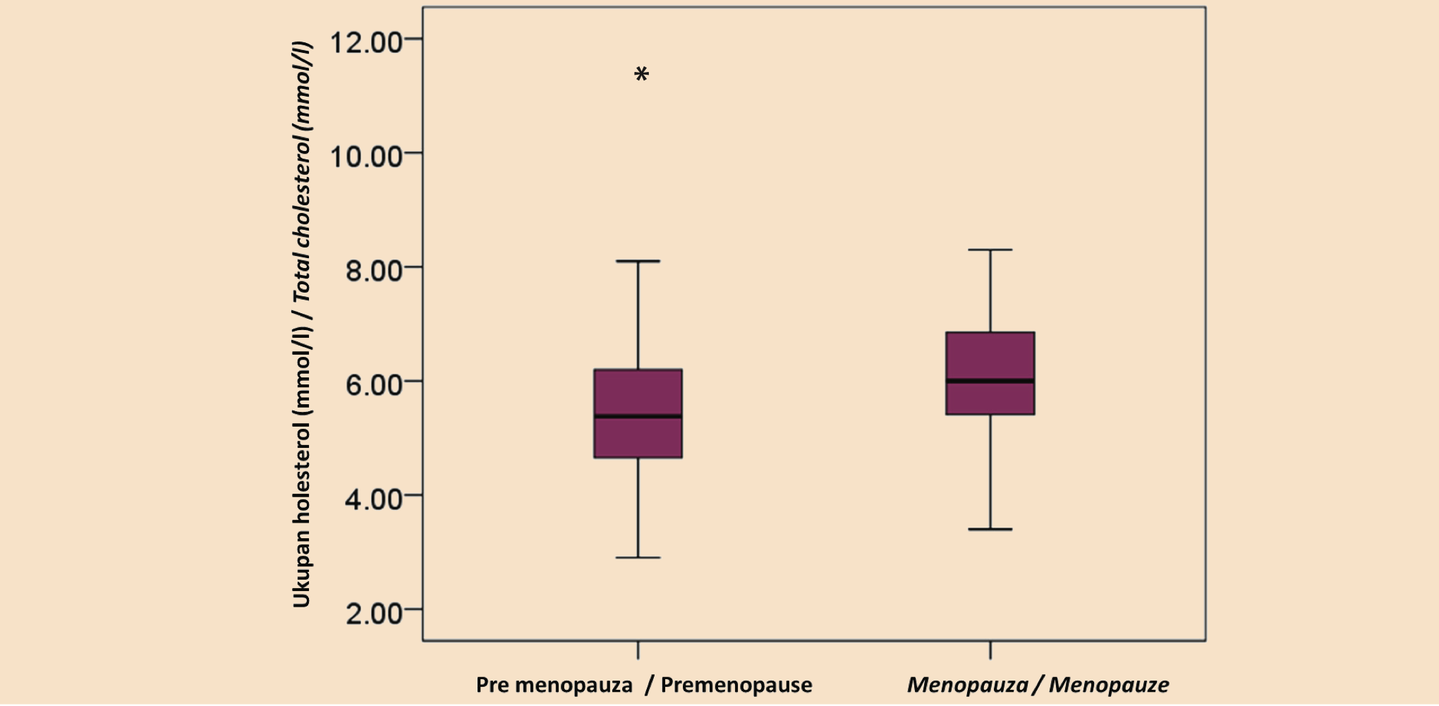
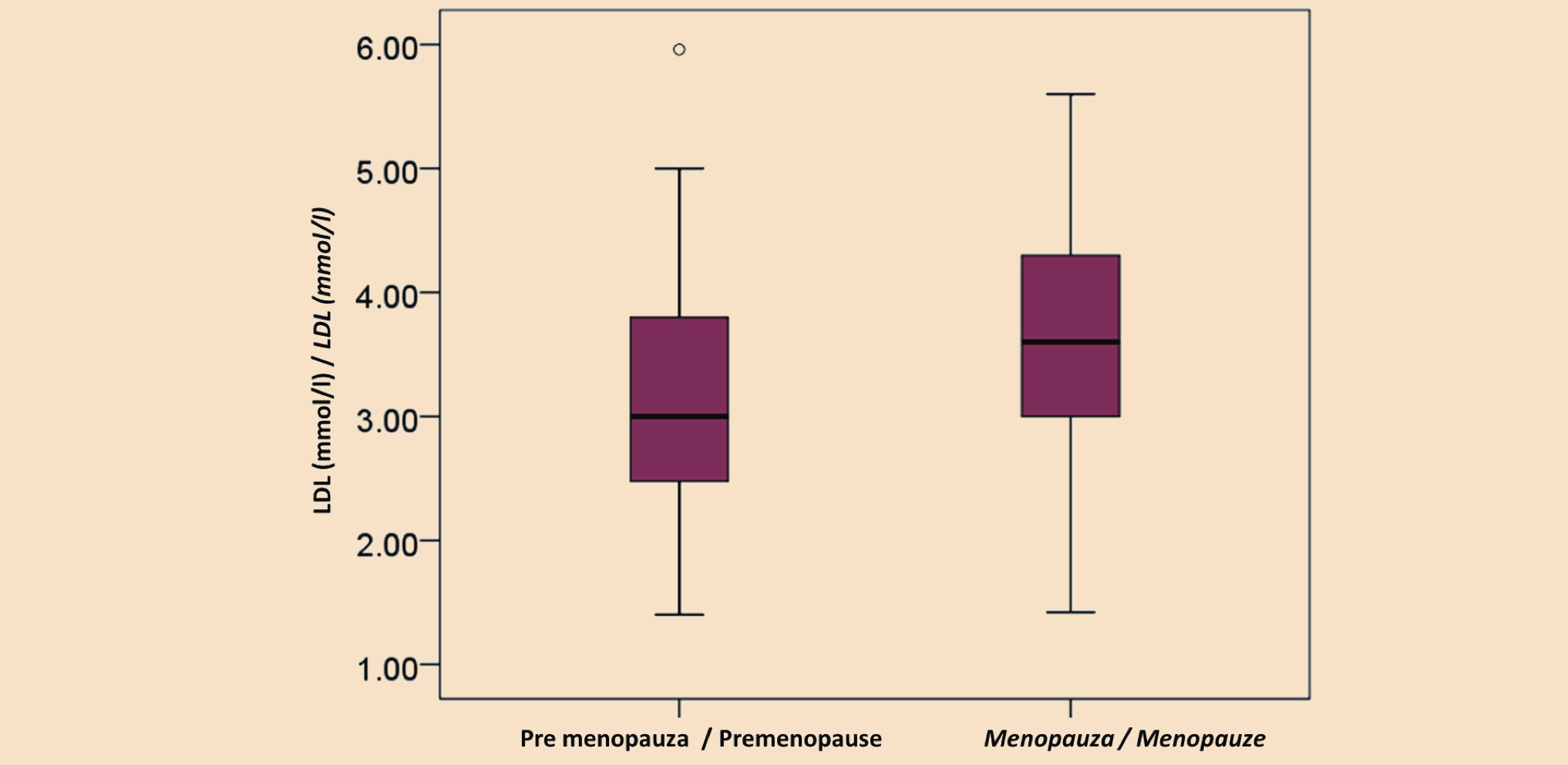
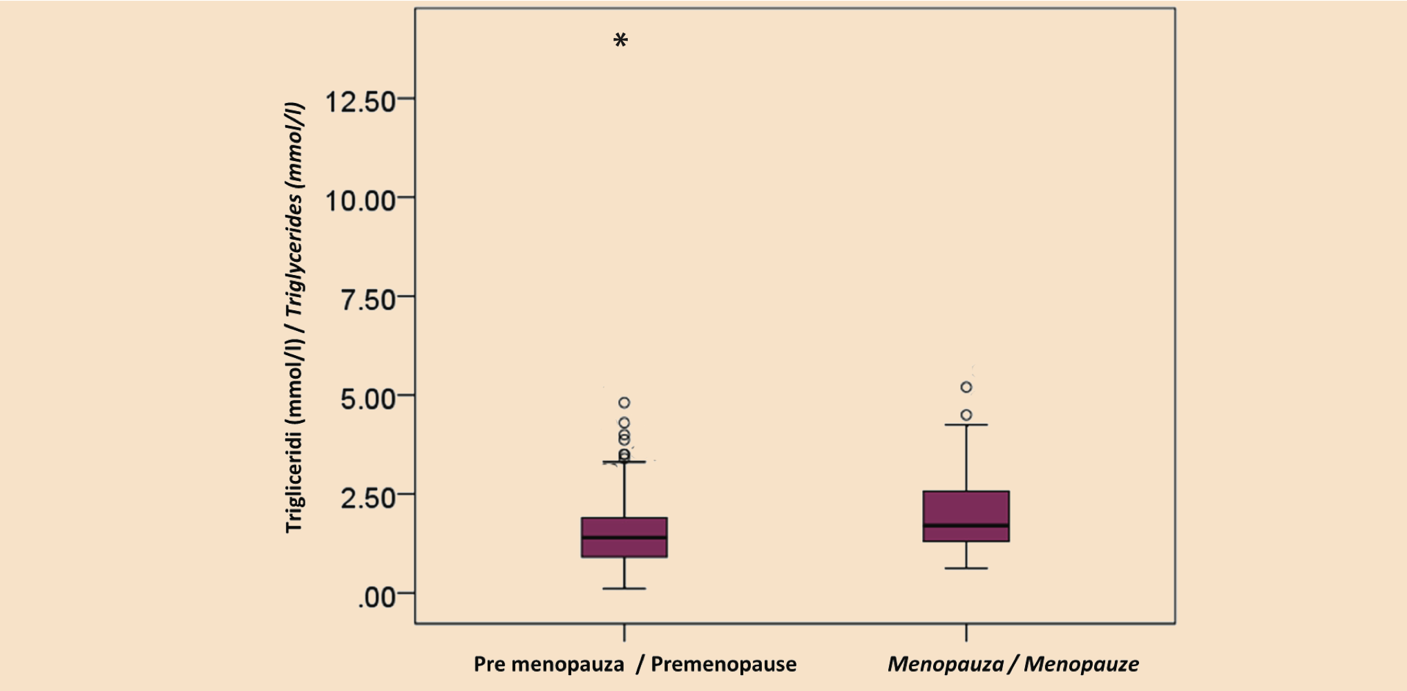
Dyslipidemia was assessed by measuring total cholesterol, LDL cholesterol, HDL cholesterol, and triglycerides. Women in menopause had elevated total and LDL cholesterol levels, while HDL cholesterol levels did not differ significantly. Total cholesterol in premenopausal women was 5.4 mmol/L (range 2.9–11.4), and in menopausal women, 6.0 mmol/L (range 3.4–8.3) (p < 0.001). LDL cholesterol levels in premenopausal women were 3.0 mmol/L (range 1.4–6.0), and in menopausal women, 3.6 mmol/L (range 1.4–5.6) (p = 0.001). Triglyceride levels were significantly higher in menopausal women, amounting to 1.7 mmol/L (range 0.6– 5.2), compared to 1.4 mmol/L (range 0.1–14.0) in premenopausal women (p = 0.002) (Table 3).
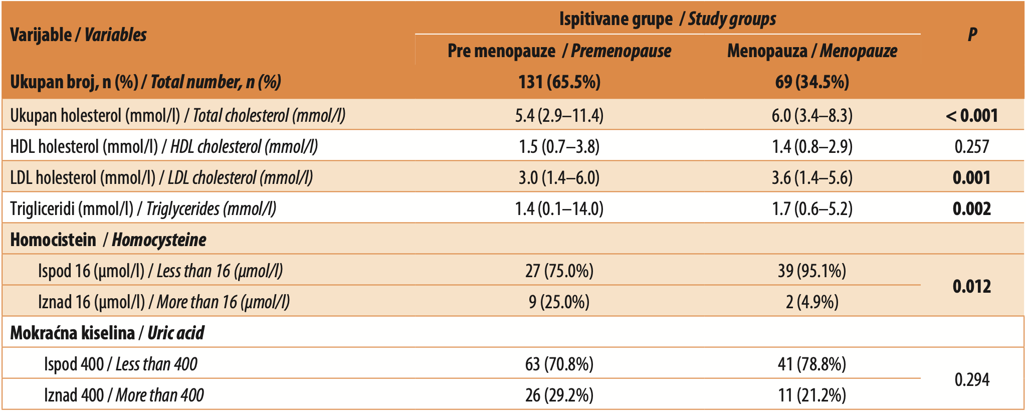
Elevated homocysteine levels (above 16 μmol/L) were significantly more frequent in premenopausal women 9 (25.0%) compared to menopausal women 2 (4.9%) (p = 0.012).
DISCUSSION
The prevalence of increased body weight and obesity continues to rise, with women being more represented than men in the obese population. This is supported by recent studies on the prevalence of obesity among individuals older than 60 years, showing rates of 37.5% in men and 39.4% in women [1]. Changes in reproductive hormones during menopause and the menopausal transition contribute to accelerated weight gain, a phenomenon that is particularly pronounced during the menopausal transition and in the first few years after the last menstruation. Thus, the higher prevalence of type 2 diabetes mellitus in women may be a consequence of obesity. Inflammation in atherogenesis, associated with obesity and diabetes, increases cardiovascular risk. Women are more likely to progress from prediabetes to type 2 diabetes mellitus [9],[14].
In the original Framingham cohort, an association between glycated hemoglobin (HbA1c) levels and cardiovascular disease was observed in women but not in men. For each 1% increase in HbA1c, the relative odds of cardiovascular disease increased by 1.39 times in women. In a meta-analysis, the rate of fatal coronary disease was higher among patients with diabetes compared to those without diabetes (5.4% vs. 1.6%) [14]. Early diagnosis of diabetes is essential, particularly for preventing microvascular complications.
Hypertension may result from vascular endothelial dysfunction and an imbalance of endothelial mediators (endothelin, nitric oxide, prostacyclin) that regulate vascular function. Estrogens induce nitric oxide (NO) synthesis and increase the formation of inositol 1,4,5-trisphosphate (IP3), which stimulates the release of Ca²+from the endoplasmic reticulum. Ca²+ forms a complex with calmodulin, which then binds and initiates activation of endothelial nitric oxide synthase (eNOS). Activated eNOS promotes the conversion of L-arginine to L-citrulline and the production of NO, which induces relaxation of vascular smooth muscle. The potential effects of estrogen on the vascular endothelium are evident. Increased vascular tone leads to hypertension, which in turn causes vascular damage [10]. Estrogens promote vasodilation and may attenuate hypertension. They may also reduce vascular permeability, reactive oxygen species (ROS), and inflammatory cell infiltration. Estrogens may also slow the progression of atherosclerosis by improving lipid profile and reducing thromboembolism by decreasing platelet aggregation. Oxidative stress and increased production of reactive oxygen species likely represent a common molecular basis linking immunoinflammation with hypertension, which is more frequent in menopausal women due to decreased estrogen levels.
Dyslipidemia is a well-known risk factor with a direct relationship between LDL cholesterol and atherosclerotic cardiovascular disease. Studies including women have shown that reduction of LDL cholesterol is associated with reduced cardiovascular risk in both primary and secondary prevention. INTERHEART was a large case-control study that aimed to quantify the risks associated with various cardiovascular disease risk factors. Smoking and abnormal lipid profile (defined as an elevated ApoB/ApoA1 ratio) were the two strongest risk factors for myocardial infarction [7],[15]. Statin therapy has benefits in reducing cardiovascular risk; however, older women use statins 20% less frequently than men, which may be due to less frequent prescribing or a higher prevalence of statin-associated myalgia [16]. Cholesterol levels may vary throughout the life course, from young adulthood to pregnancy and the menopausal transition, and treatment should be determined accordingly. Women are at increased risk of dyslipidemia during menopause, characterized by higher total cholesterol, triglycerides, and LDL cholesterol, and lower HDL cholesterol. High levels of non-HDL cholesterol and triglycerides are more important risk factors for cardiovascular disease in women than in men, especially in women with diabetes [17]. Elevated lipoprotein(a) levels are considered a risk factor with prothrombotic, proinflammatory, and proatherogenic effects in both sexes.
Hypertension, diabetes, physical inactivity, and alcohol use were more strongly associated with myocardial infarction in women than in men, as demonstrated by the INTERHEART study [7]. The Prospective Multicenter Imaging Study for Evaluation of Chest Pain (PROMISE) showed that women with stable symptoms of coronary disease had a higher burden of risk factors compared to men [8].
Increased body mass index (BMI) is associated with increased cardiovascular risk in both sexes; however, there are sex differences in fat distribution, including visceral and subcutaneous fat [18]. Women predominantly accumulate subcutaneous fat, whereas men accumulate more visceral fat. During menopause, women experience increased visceral and intramuscular fat infiltration compared with premenopausal women of similar age, contributing to insulin resistance and inflammation [1],[19]. Certain cardiovascular risk factors are associated with increased fat mass and obesity. Hypertension is associated with overweight and obesity in women, while diabetes is associated with abdominal obesity in women [18]. Weight reduction is associated with lower cholesterol levels, lower blood pressure, and a reduced risk of developing type 2 diabetes mellitus.
The results of our study are consistent with findings from European studies showing that the prevalence of metabolic disorders and hypertension significantly increases after menopause. In addition, lipid profile analyses indicate significantly higher levels of total and LDL cholesterol in postmenopausal women than in premenopausal women, further confirming unfavorable metabolic changes during this period of life [20].
In the group of perimenopausal women, hypothyroidism was significantly more frequently observed, with Hashimoto’s thyroiditis being the most common cause. In addition to improved diagnostic capabilities, the higher number of confirmed cases of hypothyroidism may be related to chronic stress and immune system disturbances associated with modern lifestyles. Environmental endocrine disruptors (found in plastics, cosmetics, and pesticides) and micronutrient deficiencies may contribute to the rising incidence of hypothyroidism [21]. Women are particularly susceptible due to the influence of estrogen on immune response, and hormonal fluctuations may trigger autoimmune disease in genetically predisposed individuals [22].
The presence of estrogen receptors (ERα and ERβ) in large numbers in visceral adipose tissue [18] supports the direct role of estrogen in modulating visceral fat function. Estrogen inhibits lipid accumulation in adipocytes through two mechanisms: directly, by reducing lipogenesis via decreased activity of lipoprotein lipase (LPL), and indirectly, by influencing lipolysis through enhancement of hormone-sensitive lipase (HSL) activity [23]. Estrogen also indirectly regulates leptin secretion from adipose tissue and affects food intake and energy expenditure, thereby influencing body weight [1],[18].
A limitation of this study is its relatively small sample size, which may affect statistical power and the generalizability of the results. For this reason, multivariate analyses were not performed. Future studies with larger sample sizes may allow the application of more complex statistical methods and further validation of these findings.
CONCLUSION
Women, like men, have traditional modifiable cardiovascular risk factors; however, in addition, they have sex-specific factors that significantly increase cardiovascular risk. There is sexual dimorphism in body fat distribution, supported by evidence of the regulatory role of estrogen, particularly 17β-estradiol, in metabolism and regional fat distribution. A tendency toward android fat distribution is observed in older women and is likely responsible for a cascade of chronic health disorders, including hypertension, type 2 diabetes mellitus, and major cardiovascular events. These findings are important for the healthcare system, as body weight control at all ages, particularly in menopausal women, should be a priority. Experimental data and clinical studies indicate complex differences in the regulation of vasomotor function of microvascular blood vessels between women and men, contributing to the understanding of sex differences and the specific risk of ischemic heart disease in women. A better understanding of pathophysiological mechanisms may help plan new strategies tailored to women for the prevention, detection, and treatment of cardiometabolic diseases.
Notification: TThis paper was previously presented at the 34th European Meeting on Hypertension and Cardiovascular Protection, May 23–26, 2025, Milan, Italy. The abstract was published in the Journal of Hypertension (2025), Vol. 43, Suppl. 1.
Informations

-
Keywords:
-
Received:
-
Revised:
-
Accepted:
-
Online first:
-
DOI:
-
Cite this article:

Sonja Smiljić
Faculty of Medicine, University of Priština – Kosovska Mitrovica
bb Anri Dinana Street, 38220 Kosovska Mitrovica, Serbia
E-mail:
1. Rehman A, Lathief S, Charoenngam N, Pal L. Aging and Adiposity-Focus on Biological Females at Midlife and Beyond. Int J Mol Sci. 2024 Mar 4;25(5):2972. doi: 10.3390/ijms25052972. [CROSSREF]
2. Shifren JL, Gass ML; NAMS Recommendations for Clinical Care of Midlife Women Working Group. The North American Menopause Society recommendations for clinical care of midlife women. Menopause. 2014 Oct;21(10):1038-62. doi: 10.1097/GME.0000000000000319. [CROSSREF]
3. Elder P, Sharma G, Gulati M, Michos ED. Identification of female-specific risk enhancers throughout the lifespan of women to improve cardiovascular disease prevention. Am J Prev Cardiol. 2020 Jun 6;2:100028. doi: 10.1016/j.ajpc.2020.100028. [CROSSREF]
4. Peters SA, Huxley RR, Woodward M. Diabetes as risk factor for incident coronary heart disease in women compared with men: a systematic review and meta-analysis of 64 cohorts including 858,507 individuals and 28,203 coronary events. Diabetologia. 2014 Aug;57(8):1542-51. doi: 10.1007/s00125-014-3260-6. [CROSSREF]
5. Nguyen AH, Hurwitz M, Sullivan SA, Saad A, Kennedy JLW, Sharma G. Update on sex specific risk factors in cardiovascular disease. Front Cardiovasc Med. 2024 Feb 6;11:1352675. doi: 10.3389/fcvm.2024.1352675. [CROSSREF]
6. Huxley RR, Woodward M. Cigarette smoking as a risk factor for coronary heart disease in women compared with men: a systematic review and meta-analysis of prospective cohort studies. Lancet. 2011 Oct 8;378(9799):1297- 305. doi: 10.1016/S0140-6736(11)60781-2. [CROSSREF]
7. Anand SS, Islam S, Rosengren A, Franzosi MG, Steyn K, Yusufali AH, et al.; INTERHEART Investigators. Risk factors for myocardial infarction in women and men: insights from the INTERHEART study. Eur Heart J. 2008 Apr;29(7):932- 40. doi: 10.1093/eurheartj/ehn018. [CROSSREF]
8. Hemal K, Pagidipati NJ, Coles A, Dolor RJ, Mark DB, Pellikka PA, et al. Sex Differences in Demographics, Risk Factors, Presentation, and Noninvasive Testing in Stable Outpatients With Suspected Coronary Artery Disease: Insights From the PROMISE Trial. JACC Cardiovasc Imaging. 2016 Apr;9(4):337-46. doi: 10.1016/j.jcmg.2016.02.001. [CROSSREF]
9. Kalyani RR, Lazo M, Ouyang P, Turkbey E, Chevalier K, Brancati F, et al. Sex differences in diabetes and risk of incident coronary artery disease in healthy young and middle-aged adults. Diabetes Care. 2014;37(3):830-8. doi: 10.2337/dc13-1755. [CROSSREF]
10. Smiljić S, Ilić A. The impact of dyslipidemia and hyperhomocysteinemia on the development of arterial hypertension. Galenika Medical Journal. 2024;3(9):7-13. doi: 10.5937/Galmed2409007S. [CROSSREF]
11. Joyner JM, Hutley LJ, Cameron DP. Estrogen receptors in human preadipocytes. Endocrine. 2001 Jul;15(2):225-30. doi: 10.1385/ENDO:15:2:225. [CROSSREF]
12. Mou H, Zhang J, Guo Y, Xu L, Luo X. Effects of key physiological parameters on cardiovascular disease and osteoporosis risk in perimenopausal and postmenopausal women. Sci Rep. 2025 Jan 22;15(1):2814. doi: 10.1038/s41598- 025-86613-8. Erratum in: Sci Rep. 2025 Mar 25;15(1):10251. doi: 10.1038/ s41598-025-95430-y. [CROSSREF]
13. Nie G, Yang X, Wang Y, Liang W, Li X, Luo Q, et al. The Effects of Menopause Hormone Therapy on Lipid Profile in Postmenopausal Women: A Systematic Review and Meta-Analysis. Front Pharmacol. 2022 Apr 12;13:850815. doi: 10.3389/fphar.2022.850815. [CROSSREF]
14. Mahabadi AA, Massaro JM, Rosito GA, Levy D, Murabito JM, Wolf PA, et al. Association of pericardial fat, intrathoracic fat, and visceral abdominal fat with cardiovascular disease burden: the Framingham Heart Study. Eur Heart J. 2009 Apr;30(7):850-6. doi: 10.1093/eurheartj/ehn573. [CROSSREF]
15. Yusuf S, Hawken S, Ounpuu S, Dans T, Avezum A, Lanas F, et al.; INTERHEART Study Investigators. Effect of potentially modifiable risk factors associated with myocardial infarction in 52 countries (the INTERHEART study): case-control study. Lancet. 2004 Sep 11-17;364(9438):937-52. doi: 10.1016/S0140-6736(04)17018-9. [CROSSREF]
16. Kostis WJ, Cheng JQ, Dobrzynski JM, Cabrera J, Kostis JB. Meta-analysis of statin effects in women versus men. J Am Coll Cardiol. 2012 Feb 7;59(6):572- 82. doi: 10.1016/j.jacc.2011.09.067. Erratum in: J Am Coll Cardiol. 2012 Apr 17;59(16):1491.
17. Lu W, Resnick HE, Jablonski KA, Jones KL, Jain AK, Howard WJ, et al. NonHDL cholesterol as a predictor of cardiovascular disease in type 2 diabetes: the strong heart study. Diabetes Care. 2003 Jan;26(1):16-23. doi: 10.2337/diacare.26.1.16. [CROSSREF]
18. Welsh A, Hammad M, Piña IL, Kulinski J. Obesity and cardiovascular health. Eur J Prev Cardiol. 2024 Jun 3;31(8):1026-35. doi: 10.1093/eurjpc/zwae025. [CROSSREF]
19. Nilsson S, Qvick A, Henriksson M, Lawesson SS, Holm AS, Leander K. Menopausal Vasomotor Symptoms and Subclinical Atherosclerotic Cardiovascular Disease: A Population-Based Study. J Am Heart Assoc. 2024 Sep 3;13(17):e033648. doi: 10.1161/JAHA.123.033648. [CROSSREF]
20. Marchi R, Dell’Agnolo CM, Lopes TCR, Gravena AAF, Demitto MO, Brischiliari SCR, et al. Prevalence of metabolic syndrome in pre- and postmenopausal women. Arch Endocrinol Metab. 2017 Mar-Apr;61(2):160-6. doi: 10.1590/2359-3997000000253. [CROSSREF]
21. Wyne KL, Nair L, Schneiderman CP, Pinsky B, Antunez Flores O, Guo D, et al. Hypothyroidism Prevalence in the United States: A Retrospective Study Combining National Health and Nutrition Examination Survey and Claims Data, 2009-2019. J Endocr Soc. 2022 Nov 10;7(1):bvac172. doi: 10.1210/jendso/bvac172. [CROSSREF]
22. Vargas-Uricoechea H, Castellanos-Pinedo A, Urrego-Noguera K, Pinzón-Fernández MV, Meza-Cabrera IA, Vargas-Sierra H. A Scoping Review on the Prevalence of Hashimoto’s Thyroiditis and the Possible Associated Factors. Med Sci (Basel). 2025 Apr 10;13(2):43. doi: 10.3390/medsci13020043. [CROSSREF]
23. Pant A, Chew DP, Mamas MA, Zaman S. Cardiovascular Disease and the Mediterranean Diet: Insights into Sex-Specific Responses. Nutrients. 2024 Feb 19;16(4):570. doi: 10.3390/nu16040570. [CROSSREF]
- Table 1. Cardiovascular risk factors in premenopausal and menopausal women
- Table 2. Prevalence of comorbidities in patients before menopause and in menopause
- Table 3. Biochemical variables in premenopausal and menopausal women
- Graph 1. Total cholesterol levels in premenopausal and menopausal women
- Graph 2. LDL cholesterol levels in premenopausal and menopausal women
- Graph 3. Тriglyceride levels in premenopausal and menopausal women