


Surgical treatment of radiation-induced anterior chest wall osteosarcoma after breast carcinoma: a case report
- Military Medical Academy, Clinic for Chest and Cardiosurgery, Belgrade, Serbia
- University of Defense, Military Medical Academy, Medical Faculty, Belgrade, Serbia
- Military Medical Academy, Clinic for Plastic Surgery and Burns, Belgrade, Serbia
ABSTRACT
Introduction/Objective: Radiotherapy for breast cancer can rarely lead to radiation-induced osteosarcoma (RIOS), a highly aggressive malignancy. This article aims to present the specificity of the surgical treatment applied and the level of aggressiveness of this rare malignant tumor.
Case report: A 63-year-old female patient was admitted to the Chest Surgery Department of the Military Medical Academy due to a large exulcerated sternal mass releasing a purulent secretion, initially misdiagnosed as recurrent breast carcinoma 15 years post-treatment, and anemia. Biopsy confirmed high-grade osteosarcoma following prior radiotherapy. After metastasis was ruled out, the patient received neoadjuvant chemotherapy and underwent radical resection of the sarcoma, sternum, anterior III–VII ribs, and adjacent soft tissues. Chest wall reconstruction was achieved using titanium bars, clips, and a latissimus dorsi myocutaneous flap. Upon local recurrence, further resection of the manubrium and anterior II ribs, with soft tissues, was performed. A year after the initial operation, a suspicious subcutaneous mass in the left pectoral region was excised for histopathological analysis.
Conclusion: RIOS of the chest wall exhibits a more aggressive course and worse prognosis than sporadic chest wall sarcomas, with higher rates of recurrence despite wide resection, as demonstrated in this case. Reconstruction with titanium bars and clips, after extensive anterior chest wall resection, provides a rigid and feasible alternative for restoring stability. This case underscores the importance of recognizing RIOS as a rare but serious late complication of breast cancer radiotherapy and highlights the complexity and specificity of surgical management and reconstruction for optimal outcomes.
INTRODUCTION
Osteosarcomas account for 10%–15% of all primary malignant tumors of the chest wall, which, in turn, account for only 1%–2% of all primary tumors [1]. The association between radiotherapy (RT) applied as a part of breast cancer oncological treatment and the occurrence of radiation-induced osteosarcoma (RIOS) in the radiation region, although extremely rare, has been previously described. These osteosarcomas have a significantly higher malignant potential and are rarer than sporadic osteosarcomas. Surgery is still the treatment of first choice in the multimodal treatment of these sarcomas.
CASE REPORT
A 63-year-old female patient was admitted to the Clinic for Thoracic Surgery of the Military Medical Academy due to a large exulcerated sternal mass with purulent discharge and anemia. The patient had received oncological treatment for breast cancer 15 years earlier. She first underwent bilateral mastectomy, followed by adjuvant chemotherapy (CHT) (four cycles of Adriamycin–Cyclophosphamide and four cycles of Taxotere) and radiotherapy. Finally, the tumor board recommended hormonal therapy (tamoxifen) for a period of five years.
During routine follow-up, a multi-slice computed tomography (MSCT) chest scan revealed an osteolytic tumor lesion measuring five centimeters in the region of the sternum, suggestive of metastatic breast carcinoma, along with described multiple nonspecific micronodular changes in the lungs. Bone scintigraphy showed that the described lesion involved the distal portions of the sternal body and the xiphoid process and demonstrated increased radiotracer uptake.
The oncological tumor board initially interpreted the lesion as a relapse of the primary carcinoma, and made a decision that treatment should be continued with hormonal therapy.
The chest MSCT performed five months after the initial scan that had initially detected the lesion, showed a progression, with the tumor measuring 5 cm × 5 cm × 8.5 cm in total. The patient was therefore referred for biopsy of the tumor (pathohistological (PH) finding: inconclusive). A rebiopsy was performed one month later, and pathohistological analysis confirmed osteosarcoma. Based on the decision made by the tumor board, chemotherapy according to the adriamycin–ifosfamide protocol was indicated and administered (a total of four cycles).
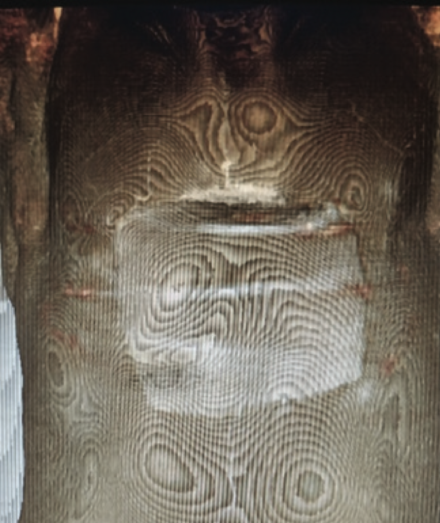
The follow-up MSCT of the chest and abdomen performed four months after pathohistological (PH) verification and immediately after the last cycle of chemotherapy, revealed a large expansive tumor mass of the body of the sternum, with larger calcifications and a cystic component causing bone destruction, exulcerating the skin, and propagating retrosternally with compression of the right atrium and displacement of the mediastinum to the left (Image 1). In the pulmonary parenchyma there were no focal lesions; the mediastinum showed no lymphadenopathy; the abdominal findings were unremarkable.
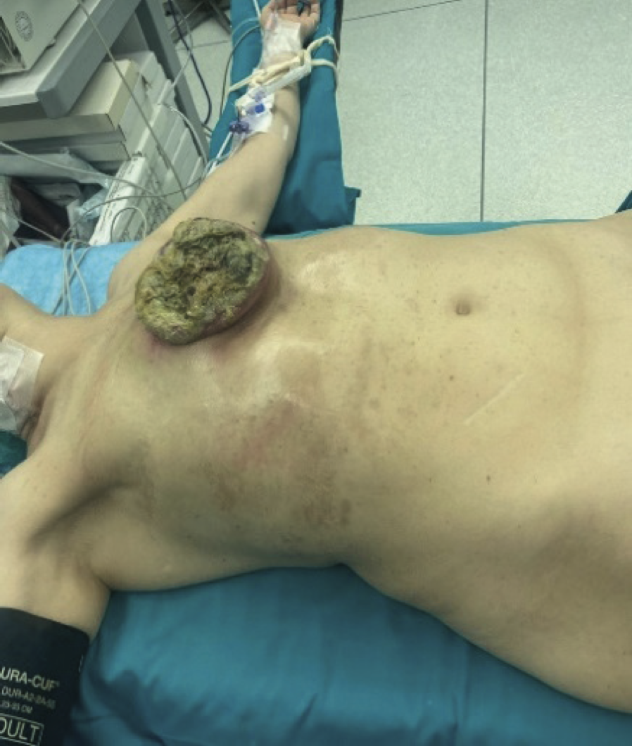
Through a multidisciplinary approach, after presentation to the tumor board, a decision was made to proceed with surgical treatment – a wide resection of the osteosarcoma of the anterior chest wall with rigid and soft-tissue reconstruction of the defect.
As part of preoperative preparation, imaging of the thoracodorsal arteries was performed using color Doppler ultrasonography. The findings were normal – both arteries showed physiological flow, and the accompanying veins had no signs of thrombosis.
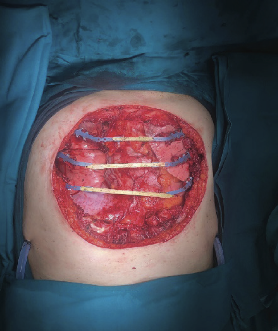
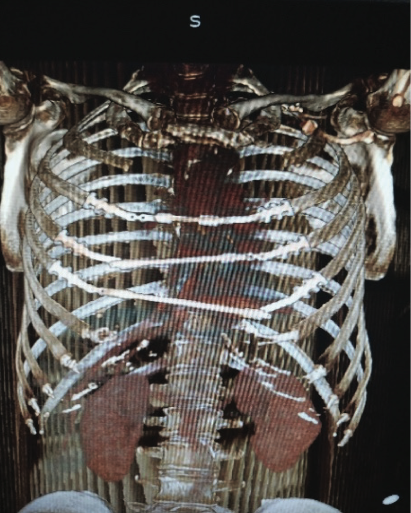
An en bloc resection of the osteosarcoma was performed together with the greater part of the body of the sternum and the xiphoid process, with resection of the anterior portions of the third through seventh ribs bilaterally up to four centimeters from the margins of the tumor mass, along with the adjacent soft-tissue structures of the anterior chest wall and resection of the pericardium. Titanium clips and bars were used for rigid reconstruction. The concept of reconstruction was not to simulate replacement with structures resembling the sternum but rather to connect the ribs horizontally, with bars bridging the parallel ribs. Clips were fixed to the parallel ribs III, IV, and V on the right and left sides, and bridging bars were placed connecting the clips on the aforementioned ribs (Figure 2). With the help of specially designed instruments, as well as the clips themselves, it is possible to manipulate a wide range of different angles in all three axes and achieve the most stable and optimal three-dimensional configuration.
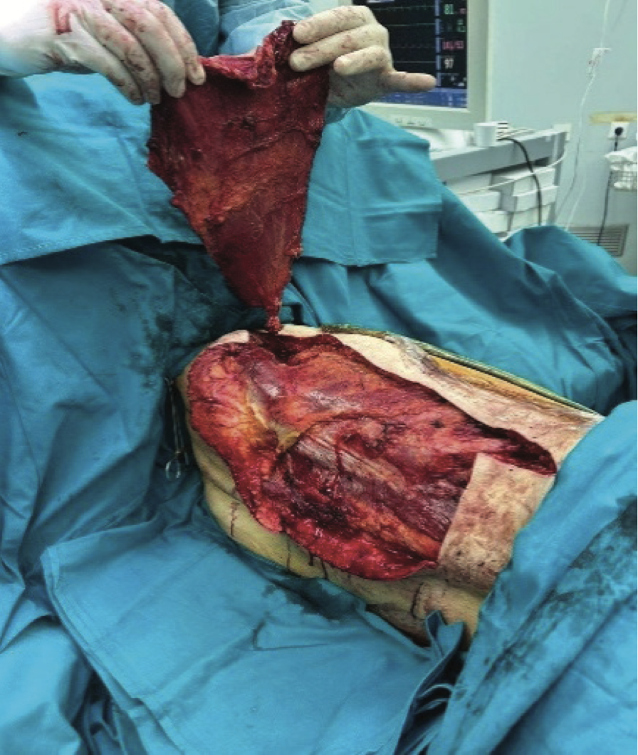
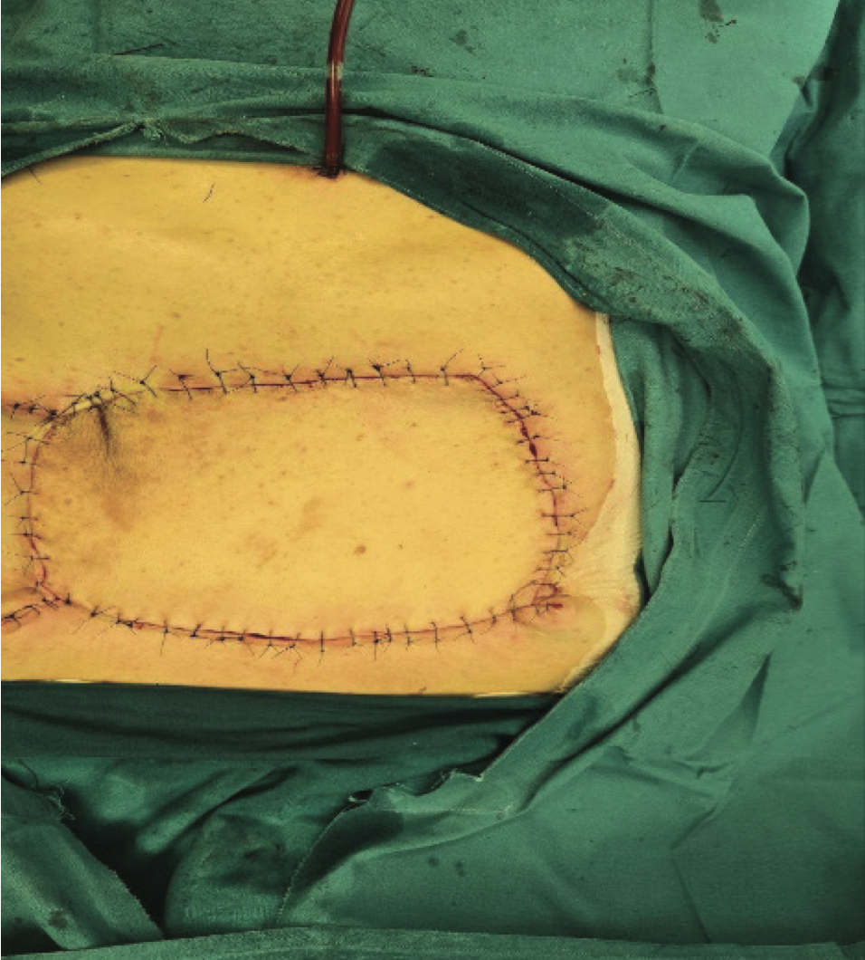
After osteosynthesis, reconstruction of the defect was performed using a myocutaneous flap of the latissimus dorsi muscle, while the secondary defect on the back was partially closed directly, sewn in layers, and partially reconstructed using a previously harvested Thiersch skin graft from the region of the right thigh.
The patient was extubated early and generally tolerated the intervention well. After drainage was reduced, the bilateral pleural drains, as well as the aspiration drain for control of bleeding from the myocutaneous flap, were removed. The wounds healed per primam, with adequate acceptance of the graft and myocutaneous flap and daily dressing. The patient was discharged for further home treatment on the twentieth postoperative day without significant complications.
The following PH finding was obtained: solitary tumor measuring 220 mm × 130 mm, high-grade osteosarcoma (G3) in pT2 stage (WHO Classification of Bone Tumors, 5th Edition). Tumor tissue was present at the sternal resection margin (R1); no tumor tissue was present in the other structures. Immunophenotype of the tumor cells: Vimentin +, SATB +/−, SMA +/−, CD31 −, CD34 −, EMA −, S100 −/+.
In keeping with the tumor board decision, three months after the operation, treatment was continued with three cycles of chemotherapy according to the adriamycin – ifosfamide protocol. Follow-up MSCT imaging of the chest, six months after surgery and one and a half months after the last administered cycle of chemotherapy, showed the following finding: presence of the residual/recurrent previously described tumor lesion of the proximal part of the sternum measuring 45 mm × 62 mm.
The tumor board again indicated surgical treatment, and seven months after the first operation a partial resection of the distal part of the manubrium of the sternum was performed with en bloc resection of the anterior portions of the second ribs bilaterally and wedge resection of the portion of the left lung adherent to the tumor, i.e., subtotal resection of the sternum with preservation of the sternoclavicular and sternocostal connections with wedge resection of the left lung. In the second operation there was no need for additional reconstruction.
The following PH finding was obtained: Osteosarcoma “high-grade” in textu ossealis costae infiltrativum. No tumor tissue was present at the sternal resection margin (R0). Tumor cells were present in the lateral soft-tissue structures of the chest wall and in the lungs − R1 resection.
MSCT of the chest, three months after the second operation, detected three oval-shaped, soft-tissue lesions, partially calcified, with central hypodense zones suggestive of necrosis. The first lesion was positioned mediastinally, para-aortically, at position 6. The second was on the right side, anteriorly, supradiaphragmally, adjacent to the diaphragmatic pleura and posterior contour of the anterior chest wall. The third lesion was positioned paramediastinally, adjacent to the superior vena cava. All three lesions measured approximately two centimeters in diameter, primarily corresponding to residual/recurrent disease or metastases of the treated neoplastic lesion.
The tumor board indicated cyclophosphamide monotherapy for the duration of one month.
Less than a year after the first operation, clinical examination verified a tumor lesion in the subcutaneous tissue of the left pectoral region, which was ovalshaped, approximately four centimeters in diameter, painless, mobile relative to the underlying tissue, suspect of recurrence. The lesion was therefore removed for PH verification. It was decided that the other lesions would not be resected at that time.
A year after the initial surgery, the patient is in good general condition, motivated for further multidisciplinary evaluation and selection of the most effective, but also the most rational therapeutic approach.
DISCUSSION
Osteosarcomas of the chest wall have a significantly poorer prognosis than sarcomas of other regions, with a five-year survival rate of 15%–20% [1]. Radiation-induced osteosarcomas (RIOS) have an even worse prognosis than sporadic osteosarcomas; they are characterized by high grade, significant mitotic activity, a more aggressive course, and poorer prognosis [2],[3]. Whenever possible, tumors of the chest wall should undergo preoperative biopsy. Needle or core biopsy is generally sufficient. Incisional biopsy may be performed to obtain a larger sample in rare tumors.
In our patient, the lesion was identified and monitored on consecutive contrast-enhanced CT scans of the chest and abdomen. The introduction of PET-CT imaging into standard preoperative preparation, along with magnetic resonance imaging of the head, might better select patients, potentially better demarcate the tumor, and ultimately provide better surgical outcomes.
After pathohistological confirmation, multidisciplinary assessment, and induction chemotherapy, surgery is the treatment of choice when feasible [4]. The greatest challenge in the surgical treatment of this disease is planning and performing reconstruction of the defect created by radical resection of such a tumor. Historically, reconstruction of the resected sternum has always been more difficult than reconstruction of other bony structures. However, with the introduction of the technique that uses titanium clips and bars, it has become feasible [5]. Titanium has exceptional strength, it is not prone to frequent infections, and when the described technique is applied it can mimic the normal movements of the chest wall [6],[7].
In the literature, the expert consensus is that for high-grade tumors of the chest wall a distance of four centimeters from the macroscopically visible tumor is necessary. However, in radiation-induced osteosarcomas, additional caution is required because of the high rate of local recurrence (39%) and spreading of malignancy (27%) [8]. Rigid reconstruction, particularly in subtotal sternotomy, is feasible and may be considered as the initial treatment for osteosarcomas of the distal sternum when the disease is not disseminated, although each patient should be approached individually.
Transposition of the myocutaneous latissimus flap is often sufficient for the soft-tissue defect. However, if it is insufficient, transposition of a transverse rectus abdominis muscle flap can also be applied. The secondary defect on the back created after transposition of the myocutaneous latissimus flap is approximated and reduced by surrounding tissues, especially in individuals who have “excess tissue,” while the remainder is covered with a Thiersch skin autograft from the thigh region, in our case from the right side.
Postoperative recovery was uneventful, with mild pain, although in resections of bony structures the pain can be unbearable and difficult to manage with a combination of opioid and non-opioid analgesics, and may have the tendency of becoming chronic. The precise dissection of the neurovascular pedicle during the preparation of the ribs for the application of titanium clips certainly contributes to less pain. If the titanium clips were to clamp the neurovascular bundle to the ribs, this would lead to significant pain. R0 resection is mandatory for improved survival.
In our patient, in the first procedure, due to a positive sternal margin and microscopic presence of tumor cells, the operation did not achieve R0 resection, while the lateral soft-tissue margins and resected ribs achieved R0 resection. During the second operation, after recurrence, the lateral soft-tissue component did not achieve R0 resection, whereas the sternum did.
In such patients, a multidisciplinary approach is essential. In addition to surgery, induction and adjuvant chemotherapy may be effective. In our patient, chemotherapy was ineffective. In R1 resections, adjuvant radiotherapy may be attempted. However, since the osteosarcoma developed in previously irradiated tissue, the final effect and the potential benefit-to-harm ratio are issues of concern. In certain circumstances, stereotactic body radiation therapy (SBRT) may yield acceptable results [9]. In some cases, the use of SBRT as adjuvant therapy after breast cancer, or, in fact, the use of more precise radiation therapy compared with conventional methods, may potentially reduce the incidence of RIOS [10].
Informations

-
Keywords:
-
Received:
-
Revised:
-
Accepted:
-
Online first:
-
DOI:
-
Cite this article:

Stevan Čičić
Clinic for Chest and Cardiosurgery, Military Medical Academy
17 Crnotravska Street, 11000 Belgrade, Serbia
E-mail:
1. Shah AA, D’Amico TA. Primary chest wall tumors. J Am Coll Surg. 2010 Mar;210(3):360-6. doi: 10.1016/j.jamcollsurg.2009.11.012. [CROSSREF]
2. Virtanen A, Pukkala E, Auvinen A. Incidence of bone and soft tissue sarcoma after radiotherapy: a cohort study of 295,712 Finnish cancer patients. Int J Cancer. 2006 Feb 15;118(4):1017-21. doi: 10.1002/ijc.21456. [CROSSREF]
3. Bjerkehagen B, Småstuen MC, Hall KS, Skjeldal S, Smeland S, Fosså SD. Why do patients with radiation-induced sarcomas have a poor sarcoma-related survival? Br J Cancer. 2012 Jan 17;106(2):297-306. doi: 10.1038/bjc.2011.559. [CROSSREF]
4. Schwarz RE, Burt M. Radiation-associated malignant tumors of the chest wall. Ann Surg Oncol. 1996 Jul;3(4):387-92. doi: 10.1007/BF02305669. [CROSSREF]
5. Scarci M. Veritas® and STRATOSTM: An Innovative Paradigm for Chest Wall Reconstruction with a Biological Patch and Titanium Bars. CTSNet [Internet]. Chicago (IL): CTSNet; 2011 Jan 2 [Accessed: Dec 7, 2025 ]. Available from: https://www.ctsnet.org/article/veritas%C2%AE-and-stratos%E2%84%A2-innovative-paradigm-chest-wall-reconstruction-biological-patch-and [HTTP]
6. Palade E, Titu IM, Fodor L, Ciorba IM, Jentimir I, Teterea F, et al. Sternal Resections: An Attempt to Find the Ideal Reconstruction Method. Medicina (Kaunas). 2025 Apr 21;61(4):763. doi: 10.3390/medicina61040763. [CROSSREF]
7. Coonar AS, Wihlm JM, Wells FC, Qureshi N. Intermediate outcome and dynamic computerised tomography after chest wall reconstruction with the STRATOS titanium rib bridge system: video demonstration of preserved bucket-handle rib motion. Interact Cardiovasc Thorac Surg. 2011 Jan;12(1):80-1. doi: 10.1510/icvts.2010.249615. [CROSSREF]
8. Inchaustegui ML, Kon-Liao K, Ruiz-Arellanos K, Silva GAE, Gonzalez MR, Pretell-Mazzini J. Treatment and Outcomes of Radiation-Induced Soft Tissue Sarcomas of the Extremities and Trunk-A Systematic Review of the Literature. Cancers (Basel). 2023 Nov 25;15(23):5584. doi: 10.3390/cancers15235584. [CROSSREF]
9. Brown LC, Lester RA, Grams MP, Haddock MG, Olivier KR, Arndt CA, et al. Stereotactic body radiotherapy for metastatic and recurrent ewing sarcoma and osteosarcoma. Sarcoma. 2014;2014:418270. doi: 10.1155/2014/418270. [CROSSREF]
10. Pollock BE, Link MJ, Stafford SL, Parney IF, Garces YI, Foote RL. The Risk of Radiation-Induced Tumors or Malignant Transformation After Single-Fraction Intracranial Radiosurgery: Results Based on a 25-Year Experience. Int J Radiat Oncol Biol Phys. 2017 Apr 1;97(5):919-23. doi: 10.1016/j.ijrobp.2017.01.004. [CROSSREF]
- Figure 1. Exulcerated purulent-secreting tumor of the anterior chest wall before the first operation
- Figure 2. Rigid reconstruction with titanium clips and bars – first operation
- Figure 3. Raised latissimus dorsi myocutaneous flap – first operation
- Figure 4. Closed defect of the anterior chest wall – first operation
- Figure 5. Multiplanar reconstruction of soft-tissue components after the second operation
- Figure 6. Multiplanar reconstruction of rigid components after the second operation