


Ductal carcinoma in situ: current diagnostic and therapeutic approaches
- University of Defence, Military Medical Academy, Belgrade, Serbia
- University Clinical Center Niš, Clinic for Thoracic Surgery, Niš, Serbia
ABSTRACT
Advancements in breast cancer screening have significantly increased the incidence of ductal carcinoma in situ (DCIS) diagnoses. Despite its favorable prognosis, DCIS poses a clinical challenge due to its biological heterogeneity and potential progression to invasive ductal carcinoma (IDC). Mammography remains the primary diagnostic modality, with microcalcifications as the most common radiological finding. Management is primarily surgical, involving either breast-conserving surgery (BCS) or mastectomy, depending on the extent of the lesion. In patients undergoing BCS, adjuvant radiotherapy is recommended to reduce the risk of local recurrence, while hormonal therapy (tamoxifen or aromatase inhibitors) is indicated for hormone receptor-positive tumors. Axillary lymph node dissection is not routinely performed but may be considered in cases with suspected microinvasion. Recently, active surveillance has emerged as a potential alternative for selected low-risk patients. Ongoing research is focused on the identification of prognostic factors to enable more precise risk stratification.
This review provides an overview of current multidisciplinary approaches to the diagnosis and treatment of DCIS, based on the latest scientific evidence.
INTRODUCTION
Ductal carcinoma in situ (DCIS) is a non-invasive form of breast cancer characterized by the proliferation of malignant epithelial cells within the ductal system, without breaching the basement membrane or infiltrating the surrounding tissue [1]. Although it does not have metastatic potential, DCIS is considered a precursor for invasive ductal carcinoma (IDC) [1]. Studies indicate that 25–60% of untreated DCIS cases may progress to IDC [2].
With the increasing implementation of breast-conserving surgery (BCS), there is an escalating necessity for accurate stratification of lesions with a higher likelihood of recurrence or progression into IDC. To address this need, new classification systems have been developed, primarily based on nuclear grade and the presence of necrosis [3]. Several studies have confirmed the clinical relevance of this approach, demonstrating that high nuclear grade and/or necrosis (especially comedo necrosis) are associated with an increased risk of early local recurrence [4],[5].
Over the past decade, professional discussions have intensified regarding the most appropriate therapeutic approach to DCIS. While surgical resection with adjuvant radiotherapy remains the standard treatment option, an ongoing debate concerns whether certain patients (particularly those with low-grade DCIS and favorable biological characteristics) are being unnecessarily exposed to aggressive treatment modalities. Recently, active surveillance has increasingly been considered as a potential option for these patients. However, due to the need to balance the risk of progression against the potential overtreatment of indolent lesions, therapeutic protocols remain a matter of expert debate [6].
This paper aims to present current diagnostic and therapeutic approaches to DCIS, with special emphasis on the challenges of making optimal clinical decisions.
DIAGNOSIS
Anamnesis and Clinical Examination
A clinical examination is mandatory before the initiation of radiological diagnostic procedures. In the context of hereditary DCIS risk, particular attention should be given to detailed family history. Following risk assessment, patients should be referred for genetic counseling when indicated. The National Comprehensive Cancer Network (NCCN) recommends routine testing for estrogen receptor expression to determine the potential benefit of adjuvant hormonal therapy, while progesterone receptor testing remains optional [6].
Mammography
During the early phase of screening implementation, the incidence of DCIS diagnoses increased by more than 500% [7]. Currently, DCIS accounts for 20–25% of all newly diagnosed breast cancer cases [8].
Microcalcifications represent the primary mammographic manifestation of DCIS. According to Dershaw et al., mammography detected their presence in 98% of cases [9]. Similar findings were reported by Stomper et al., who particularly emphasized their distinctive appearance [10]. In their study, calcifications associated with DCIS were described as pleomorphic, typically clustered, and often arranged in linear or segmental patterns following the distribution of breast ducts. These patterns, characteristic of malignant processes, help distinguish them from calcifications related to benign changes, which are generally round and randomly distributed [9],[10].
Another key aspect of preoperative DCIS assessment is evaluating the extent of tumor microcalcifications. Although mammography can provide valuable information about their presence, it may underestimate lesion size, particularly in low- and intermediate-grade DCIS. In studies using only standard bilateral mammography in mediolateral and craniocaudal projections, lesion size was underestimated by as much as 2 cm [11]. Additionally, peritumoral inflammation and/or fibrosis may lead to misinterpretation of mammographic findings due to the presence of a periductal mass with microcalcifications, even in the absence of invasion [12]. Therefore, mammography alone is insufficient for determining whether the basement membrane has been breached, a distinction critical for differentiating DCIS from IDC. To minimize the risk of lesion underestimation and misinterpretation of peritumoral changes, the routine use of magnification or compression mammography is recommended [13].
Magnetic Resonance Imaging (MRI)
The morphological patterns of malignancy observed on MRI are similar to those seen on mammography. The most common finding is non-nodular enhancement, which may appear as clustered or linear, following the ductal distribution within the breast [13],[14].
Some studies have demonstrated that MRI sensitivity for detecting DCIS ranges from 70–100% [15],[16]. Sensitivity varies according to tumor histology, being higher in high-grade tumors. Although MRI sensitivity is high, its specificity is lower, ranging from 37–75% [17]. The low specificity and higher rate of false positives may be attributed to benign lesions that also exhibit enhancement on MRI. Some studies have shown improved specificity when enhancement kinetics are considered, as malignant tumors typically display early and rapid signal enhancement [18].
Although MRI is not routinely utilized in all DCIS patients, it can provide valuable information in the preoperative assessment of disease extent and in detecting synchronous lesions in the contralateral breast.
Ultrasound (US)
On ultrasound, DCIS typically appears as a hypoechoic lesion with an irregular shape and poorly defined margins. These lesions usually do not exhibit significant vascularization on Doppler imaging, which helps distinguish them from IDC [19]. Ultrasound can play an essential role in detecting and assessing DCIS extent when mammographic findings are inconclusive (e.g., non-calcified lesions, high breast density). Yuan et al. demonstrated that combining these two modalities increases DCIS detection sensitivity by 10–15% [20].
Biopsy
Despite advancements in diagnostic imaging, definitive diagnosis of DCIS can only be established through histopathological analysis of tissue samples obtained via biopsy. The choice of biopsy method depends on lesion characteristics, such as size, presence of microcalcifications, and palpability. Biopsy should be performed only when indicated, based on clinical and radiological findings (BI-RADS categories 4–5), and following appropriate method selection [12].
The most commonly used techniques are core needle biopsy (CNB) and vacuum-assisted biopsy (VAB), performed under mammographic, ultrasound, or MRI guidance, depending on lesion visibility [21],[22].
Stereotactic CNB is the standard approach for non-palpable microcalcifications detected via mammography, while ultrasound-guided biopsy is preferred for sonographically visible lesions [22]. When percutaneous methods are not feasible or yield inadequate samples, wire-guided open biopsy is utilized, especially for diffuse or deeply located changes. This technique allows accurate localization and excision of suspicious lesions while adhering to oncological and aesthetic principles [6].
Sentinel Lymph Node Biopsy (SLNB)
Although some authors advocate for routine SLNB in patients at increased risk of IDC, others oppose this approach due to the low incidence of axillary metastases and the high survival rates observed in patients with pure DCIS (97–99%) [23]. The NCCN recommends selective use of SLNB in patients scheduled for mastectomy or BCS in breast regions that could compromise normal axillary lymph drainage (e.g., upper outer quadrant) [6]. In such cases, SLNB should be considered during the initial surgery, as it cannot be performed after mastectomy. The gold standard for this procedure is the dual-modality method with radioactive isotope and blue-dye method [24].
THERAPY
In recent years, there has been growing interest in less invasive therapeutic approaches for DCIS. Clinical trials such as LORIS, LORD, and COMET are investigating active surveillance as an alternative to surgery for carefully selected patients with low-risk forms of the disease [25],[26],[27]. Early results from these studies suggest comparable therapeutic outcomes to conventional treatments, at least in the short term. However, approximately 20% of patients initially diagnosed with low-risk DCIS are found to have IDC upon surgical excision [28]. Therefore, active surveillance requires strict patient selection criteria and consistent follow-up.
To better individualize treatment, various prognostic tools have been developed to support risk stratification and therapeutic decision-making. Among the traditional models, the Van Nuys Prognostic Index combines tumor size, margin width, nuclear grade, and the presence of comedo necrosis to assess the risk of local recurrence and determine optimal treatment strategies [29]. The DCIS nomogram incorporates ten clinicopathological factors to provide a precise estimate of individual recurrence risk and to guide decisions regarding radiation therapy, hormonal therapy, and surgical margin adequacy [30].
With advancements in molecular oncology, genomic tests are increasingly entering clinical practice. The Oncotype DX DCIS Score quantifies the ten-year risk of recurrence following BCS without radiotherapy, based on the expression of 12 genes, thereby assisting in decisions regarding the need for radiation [31]. Similarly, DCISionRT integrates molecular biomarkers (e.g., HER2, progesterone receptors, Ki-67, p16) with clinical data to assess the potential benefit of adjuvant radiotherapy. According to some studies, this test modifies the therapeutic plan in more than 30% of patients. These tools complement traditional clinicopathological parameters and have the potential to enhance treatment personalization, particularly by helping to avoid overtreatment in low-risk cases [32].
According to NCCN guidelines, the primary treatment modalities for DCIS are [6]:
- mastectomy,
- BCS (with or without adjuvant whole-breast radiotherapy - WBRT), and
- adjuvant hormonal therapy.
Mastectomy
Studies indicate that approximately 95% of patients with DCIS undergo surgical excision, with one-third of them receiving a mastectomy [33]. The long-term risk of local recurrence after mastectomy is very low, ranging from 1–2%, compared to 3–10% among women treated with BCS and WBRT [6]. Notably, this difference in recurrence risk does not affect long-term survival (after 20 years), which remains high in both groups (97–99%) [34],[35].
The main indications for mastectomy in DCIS include: (I) multifocality and/or extensive lesion distribution (in two or more breast quadrants), (II) high nuclear grade, (III) inability to achieve clear surgical margins after BCS, (IV) age under 40 years, (V) prior breast irradiation, and (VI) contraindications to radiotherapy [6].
Modern breast surgery has evolved towards aesthetic approaches that preserve aesthetic integrity while adhering to oncological principles. Techniques such as skin-sparing mastectomy and nipple-sparing mastectomy meet these criteria and are now the gold standard when a radical, tissue-preserving intervention is required [6].
The role of post-mastectomy radiation therapy (PMRT) in DCIS remains controversial and limited, as no specific patient group has demonstrated consistent clinical benefit. Only a small subset of high-risk patients (e.g., those with extensive disease, highgrade tumors, or positive margins) may be considered for PMRT [36].
BCS with or without WBRT
Currently, approximately 60–75% of newly diagnosed DCIS cases are treated with BCS, with or without WBRT [1],[33]. According to NCCN guidelines, BCS is appropriate for unifocal lesions (limited to a single breast quadrant) when negative surgical margins of at least 2 mm can be achieved. The guidelines also recommend adjuvant WBRT whenever feasible. In some instances, such as small (≤ 2.5 cm), low-grade lesions with negative margins (usually ≥ 2 mm), or when radiation is contraindicated, BCS alone may be considered [6].
The combination of BCS and WBRT has been extensively studied, and numerous clinical trials have confirmed its efficacy and superiority over BCS alone. Table 1 presents results from six prospective randomized trials comparing DCIS and IDC recurrence rates in patients treated with BCS alone (control group) vs. BCS with adjuvant WBRT (experimental group) [37],[38],[39],[40],[41],[42]. The results consistently demonstrate that the combined approach leads to significantly lower recurrence rates, ranging from 3.0–9.3%, compared to 7.0–19.2% in the control groups.
![Table 1. Tabular presentation of prospective studies comparing the efficacy of BCS and WBRT (experimental group) versus BCS without WBRT (control group) [37-42]](/images/2025/09/072.jpg)
Some studies have also examined long-term survival among DCIS patients treated with BCS alone vs. BCS with WBRT. Survival rates remained very high (97–99%) and did not differ significantly between the two groups [41],[42]. Therefore, while adjuvant WBRT reduces the risk of local recurrence, it does not impact overall survival, which remains equally high in both treatment strategies.
Surgical Margins
Assessment of surgical margins is widely regarded as the most critical aspect of the pathological evaluation of tissue specimens following surgical excision of DCIS. To date, no professional consensus has been established on this matter, likely due to conflicting findings across various studies.
A retrospective study involving 445 patients who underwent BCS for pure DCIS demonstrated that margin width is the most significant independent predictor of local recurrence [43]. A meta-analysis published by Dunne et al. in 2009 confirmed that patients with negative surgical margins (following BCS and adjuvant WBRT) had a 64% lower risk of recurrence compared to those with positive margins [44]. The same study also found that margins < 2 mm were associated with a significantly higher risk of recurrence than margins > 2 mm, although no statistically significant difference in recurrence rates was observed between margins ≤ 5 mm and those measuring exactly 2 mm [44]. Current guidelines from the Society of Surgical Oncology (SSO), American Society for Radiation Oncology (ASTRO), and American Society of Clinical Oncology (ASCO) recommend excision with at least 2 mm margins followed by adjuvant WBRT as the optimal strategy for minimizing local recurrence rates [45].
Accelerated Partial Breast Irradiation (APBI)
APBI is a shortened form of radiotherapy that targets only the portion of the breast where the lesion was located. This approach reduces radiation exposure to healthy tissue, shortens the duration of therapy, and may help preserve patient quality of life without compromising oncologic safety [6].
According to ASTRO guidelines, candidates for APBI include patients with screen-detected DCIS measuring ≤ 2.5 cm, grade I–II, and with surgical margins ≥ 3 mm [45].
Four prospective studies have demonstrated that APBI is effective as WBRT in achieving local disease control, with a comparable toxicity profile and improved cosmetic outcomes [46],[47],[48],[49].
Radiotherapy Boost
A radiotherapy boost refers to an additional, localized dose of radiation delivered to the tumor bed, following WBRT [50]. This approach is recommended for patients with negative surgical margins but elevated biological risk factors, such as high-grade tumors, the presence of comedo necrosis, or young age. Studies have demonstrated a significant reduction in local recurrence risk by up to 27% [50].
Hormonal Therapy
Adjuvant hormonal therapy is indicated for patients with estrogen and/or progesterone-receptor-positive DCIS following wide local excision, with or without radiotherapy.
Two main classes of drugs are used for this purpose [6]:
- selective estrogen receptor modulators (tamoxifen),
- aromatase inhibitors (anastrozole)
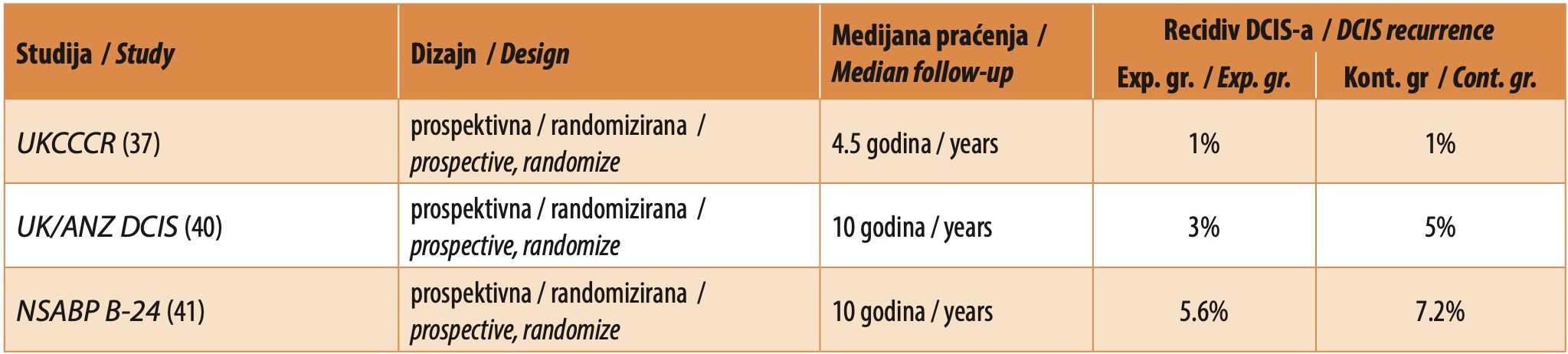
Tamoxifen is a selective estrogen receptor modulator administered at a standard dose of 20 mg per day for five years. It works by blocking estrogen receptors in breast tissue, thereby preventing hormone-induced stimulation of tumor cell proliferation [51]. Its efficacy in reducing the risk of local recurrence has been evaluated in several major clinical trials (Table 2) [37],[40],[41]. The NSABP B-24 study showed that tamoxifen, compared to placebo, reduced the 10-year DCIS recurrence rate from 7.2% to 5.6% [41]. Overall survival did not differ significantly between the groups. Similar results have been reported in other studies addressing this topic [37],[40].
 37],[40],[41]" width="2047" height="462">
Anastrozole, an aromatase inhibitor, reduces systemic estrogen levels by inhibiting the conversion of androgens to estradiol. It is typically prescribed at a dose of 1 mg daily for five years [51].
Comparative studies between tamoxifen and anastrozole, including NSABP B-35 and IBIS-II, have shown similar effectiveness in recurrence prevention (Table 3) [51],[52]. Although the overall rate of adverse events was comparable, the side effect profiles differed - fractures were more common with anastrozole. In contrast, vasomotor symptoms and thrombotic events were more frequent with tamoxifen.
 51],[52]" width="2047" height="352">
In conclusion, both tamoxifen and anastrozole play a significant role in preventing DCIS recurrence. The choice of agent should be individualized based on patient characteristics, including menopausal status and tolerance to potential side effects. Tamoxifen is the preferred option for premenopausal women, while anastrozole may be considered for postmenopausal women due to similar efficacy and a different toxicity profile.
CONCLUSION
Despite its generally favorable prognosis, DCIS requires a careful and well-considered diagnostic and therapeutic approach. The combination of BCS and adjuvant WBRT remains the most reliable strategy for reducing the risk of local recurrence, while the addition of hormonal therapy and radiotherapy boost can further improve outcomes, particularly among younger patients. In carefully selected low-risk cases, APBI may be considered an effective alternative to WBRT, offering comparable oncologic control with superior cosmetic results.
Although active surveillance has not yet been incorporated into standard clinical practice, encouraging findings from recent studies suggest that it may have a role in the management of carefully selected patients, provided that strict selection criteria are applied.
Treatment of DCIS should be individualized and guided by a combination of clinical, radiological, and pathological factors, with active patient involvement in shared decision-making.
Future research should aim to validate molecular markers that can reliably distinguish indolent from aggressive forms of DCIS, thereby enabling truly personalized treatment while preserving quality of life.
Informations

-
Keywords:
-
Received:
-
Revised:
-
Accepted:
-
Online first:
-
DOI:
-
Cite this article:

Jelena Petrović
Military Medical Academy, University of Defence
17 Crnotravska Street, 11040 Belgrade, Serbia
E-mail:
1. Hophan SL, Odnokoz O, Liu H, Luo Y, Khan S, Gradishar W, et al. Ductal carcinoma in situ of breast: from molecular etiology to therapeutic management. Endocrinology. 2022;163(4):bqac027. doi: 10.1210/endocr/bqac027. [CROSSREF]
2. Collins LC, Tamimi RM, Baer HJ, Connolly JL, Colditz GA, Schnitt SJ. Outcome of patients with ductal carcinoma in situ untreated after diagnostic biopsy: results from the Nurses’ Health Study. Cancer. 2005;103(9):1778-84. doi: 10.1002/cncr.20979. [CROSSREF]
3. Strand SH, Rivero-Gutiérrez B, Houlahan KE, Seoane JA, King LM, Risom T, et al. Molecular classification and biomarkers of clinical outcome in breast ductal carcinoma in situ: Analysis of TBCRC 038 and RAHBT cohorts. Cancer Cell. 2022;40(12):1521-36.e7. doi: 10.1016/j.ccell.2022.10.021. [CROSSREF]
4. Murata T, Yoshida M, Shiino S, Watase C, Ogawa A, Shikata S, et al. Assessment of nuclear grade-based recurrence risk classification in patients with hormone receptor-positive, human epidermal growth factor receptor 2-negative, node-positive high-risk early breast cancer. Breast Cancer. 2023;30(6):1054-64. doi: 10.1007/s12282-023-01500-2. [CROSSREF]
5. Chen J, Li Z, Han Z, Kang D, Ma J, Yi Y, et al. Prognostic value of tumor necrosis based on the evaluation of frequency in invasive breast cancer. BMC Cancer. 2023;23(1):530. doi: 10.1186/s12885-023-10943-x. [CROSSREF]
6. National Comprehensive Cancer Network. NCCN Guidelines. Ductal carcinoma in situ [Internet]. Available from: https://jnccn.org/view/journals/jnccn/20/6/article-p691.xml. Accessed: 2025 Apr 12. [HTTP]
7. White E, Lee CY, Kristal AR. Evaluation of the increase in breast cancer incidence in relation to mammography use. J Natl Cancer Inst. 1990;82(19):1546- 52. doi: 10.1093/jnci/82.19.1546. [CROSSREF]
8. Howlader N, Noone AM, Krapcho M, Miller D, Brest A, Yu M, et al. SEER cancer statistics review, 1975–2018 [Internet]. Bethesda, MD: National Cancer Institute; 2021. Available from: http://seer.cancer.gov/csr/1975_2018. Accessed: 2025 Apr 17. [HTTP]
9. Dershaw DD, Abramson A, Kinne DW. Ductal carcinoma in situ: mammographic findings and clinical implications. Radiology. 1989;170(2):411-5. doi: 10.1148/radiology.170.2.2536185. [CROSSREF]
10. Stomper PC, Connolly JL, Meyer JE, Harris JR. Clinically occult ductal carcinoma in situ detected with mammography: analysis of 100 cases with radiologic-pathologic correlation. Radiology. 1989;172(1):235-41. doi: 10.1148/ radiology.172.1.2544922. [CROSSREF]
11. Paquelet JR, Hendrick RE. Lesion size inaccuracies in digital mammography. AJR Am J Roentgenol. 2010;194(1):W115-8. doi: 10.2214/AJR.09.2927. [CROSSREF]
12. D’Alfonso TM, Ginter PS, Shin SJ. A review of inflammatory processes of the breast with a focus on diagnosis in core biopsy samples. J Pathol Transl Med. 2015;49(4):279-87. doi: 10.4132/jptm.2015.06.11. [CROSSREF]
13. Fallenberg EM, Dimitrijevic L, Diekmann F, Diekmann S, Kettritz U, Poellinger A, et al. Impact of magnification views on the characterization of microcalcifications in digital mammography. Rofo. 2014;186(3):274-80. doi: 10.1055/s0033-1350572. [CROSSREF]
14. Uslu H, Tosun M, Arslan AS. Non-mass enhancement of breast MRI: The comparison of benign and malignant pathological diagnosis and association of internal enhancement pattern and distribution with breast cancer molecular sub-types. Acta Medica Nicomedia. 2022;5(3):165–71. doi: 10.5336/actamednicommedia.2022-1128352. [CROSSREF]
15. Endrikat J, Schmidt G, Haverstock D, Weber O, Trnkova ZJ, Barkhausen J. Sensitivity of contrast-enhanced breast MRI vs X-ray mammography based on cancer histology, tumor grading, receptor status, and molecular subtype: a supplemental analysis of 2 large phase III studies. Breast Cancer (Auckl). 2022;16:11782234221092155. doi: 10.1177/11782234221092155. [CROSSREF]
16. Amitai Y, Menes TS, Scaranelo A, Fleming R, Kulkarni S, Ghai S, et al. The characteristics associated with upgrade on surgical pathology of conventional imaging occult DCIS diagnosed by MRI. Breast Cancer Res Treat. 2021;190(2):317-27. doi: 10.1007/s10549-021-06372-8. [CROSSREF]
17. Boné B, Aspelin P, Bronge L, Isberg B, Perbeck L, Veress B. Sensitivity and specificity of MR mammography with histopathological correlation in 250 breasts. Acta Radiol. 1996;37(2):208-13. doi: 10.1177/02841851960371P143. [CROSSREF]
18. Yao Y, Mou F, Kong J, Liu X. Kinetic heterogeneity improves the specificity of dynamic enhanced MRI in differentiating benign and malignant breast tumours. Acad Radiol. 2024;31(3):812-21. doi: 10.1016/j.acra.2023.10.006. [CROSSREF]
19. Cha H, Chang YW, Lee EJ, Hwang JY, Kim HJ, Lee EH, et al. Ultrasonographic features of pure ductal carcinoma in situ of the breast: correlations with pathologic features and biological markers. Ultrasonography. 2018;37(4):307- 14. doi: 10.14366/usg.17039. [CROSSREF]
20. Yuan WH, Hsu HC, Chen YY, Wu CH. Supplemental breast cancer-screening ultrasonography in women with dense breasts: a systematic review and meta-analysis. Br J Cancer. 2020;123(4):673-88. doi: 10.1038/s41416-020-0928-1. [CROSSREF]
21. Kettritz U. Modern concepts of ductal carcinoma in situ (DCIS) and its diagnosis through percutaneous biopsy. Eur Radiol. 2008;18(2):343-50. doi: 10.1007/s00330-007-0753-4. [CROSSREF]
22. Alexander N, Viljoen I, Lucas S. Stereotactic breast biopsies: radiological-pathological concordance in a South African referral unit. SA J Radiol. 2022;26(1):2463. doi: 10.4102/sajr.v26i1.2463. [CROSSREF]
23. Davey MG, O’Flaherty C, Cleere EF, Nohilly A, Phelan J, Ronane E, et al. Sentinel lymph node biopsy in patients with ductal carcinoma in situ: systematic review and meta-analysis. BJS Open. 2022;6(2):zrac022. doi: 10.1093/bjsopen/zrac022. [CROSSREF]
24. Zdravkovic D, Loboda B, Gojgic M, Toskovic B. Techniques for sentinel lymph node biopsy in breast cancer patients. Medicinska istraživanja. 2025;58(1)83- 87. doi: 10.5937/medi58-54885. [CROSSREF]
25. Francis A, Thomas J, Fallowfield L, Wallis M, Bartlett JM, Brookes C, et al. Addressing overtreatment of screen detected DCIS; the LORIS trial. Eur J Cancer. 2015;51(16):2296-303. doi: 10.1016/j.ejca.2015.07.017. [CROSSREF]
26. Schmitz RSJM, Engelhardt EG, Gerritsma MA, Sondermeijer CMT, Verschuur E, Houtzager J, et al. Active surveillance versus treatment in low-risk DCIS: Women’s preferences in the LORD-trial. Eur J Cancer. 2023;192:113276. doi: 10.1016/j.ejca.2023.113276. [CROSSREF]
27. Hwang ES, Hyslop T, Lynch T, Frank E, Pinto D, Basila D, et al. The COMET (Comparison of Operative versus Monitoring and Endocrine Therapy) trial: a phase III randomised controlled clinical trial for low-risk ductal carcinoma in situ (DCIS). BMJ Open. 2019;9(3):e026797. doi: 10.1136/bmjopen-2018-026797. [CROSSREF]
28. Chavez de Paz Villanueva C, Bonev V, Senthil M, Solomon N, Reeves ME, Garberoglio CA, et al. Factors associated with underestimation of invasive cancer in patients with ductal carcinoma in situ: precautions for active surveillance. JAMA Surg. 2017;152(11):1007-14. doi: 10.1001/jamasurg.2017.2181. [CROSSREF]
29. Silverstein MJ, Lagios MD. Treatment selection for patients with ductal carcinoma in situ (DCIS) of the breast using the University of Southern California/Van Nuys (USC/VNPI) prognostic index. Breast J. 2015;21(2):127-32. doi: 10.1111/tbj.12368. [CROSSREF]
30. Martins Maia C, Siderides C, Jaffer S, Weltz C, Cate S, Ahn S, et al. Mastectomy or margin re-excision? A nomogram for close/positive margins after lumpectomy for DCIS. Ann Surg Oncol. 2022;29(6):3740-48. doi: 10.1245/s10434-021-11293-3. [CROSSREF]
31. Solin LJ, Gray R, Baehner FL, Butler SM, Hughes LL, Yoshizawa C, et al. A multigene expression assay to predict local recurrence risk for ductal carcinoma in situ of the breast. J Natl Cancer Inst. 2013;105(10):701-10. doi: 10.1093/jnci/ djt067. [CROSSREF]
32. Bremer T, Whitworth PW, Patel R, Savala J, Barry T, Lyle S, et al. A biological signature for breast ductal carcinoma in situ to predict radiotherapy benefit and assess recurrence risk. Clin Cancer Res. 2018;24(23):5895-901. doi: 10.1158/1078-0432.CCR-18-0842. [CROSSREF]
33. Sagara Y, Mallory MA, Wong S, Aydogan F, DeSantis S, Barry WT, et al. Survival benefit of breast surgery for low-grade ductal carcinoma in situ: a population-based cohort study. JAMA Surg. 2015;150(8):739-45. doi: 10.1001/jamasurg.2015.0876. [CROSSREF]
34. Pan K, Nelson RA, Chlebowski RT, Piela R, Mullooly M, Simon MS, et al. Ductal carcinoma in situ and cause-specific mortality among younger and older postmenopausal women: the Women’s Health Initiative. Breast Cancer Res Treat. 2024;207(1):65-79. doi: 10.1007/s10549-024-07327-5. [CROSSREF]
35. Nelson RA, Lai LL, Mortimer JE, Soto Perez De Celis E, Chlebowski RT, Kruper L. Prior DCIS and overall mortality in women with stage I breast cancer. Journal of Clinical Oncology. 2021;39(15_suppl):e12593–3. doi: 10.1200/ JCO.2021.39.15_suppl.e12593. [CROSSREF]
36. Whitworth PW, Shah C, Vicini F, Rabinovitch R, Margenthaler JA, Warnberg F, et al. Identification of patients with DCIS with low-risk clinicopathology who benefit from radiation therapy with and without endocrine therapy after breast-conserving surgery assessed with the 7-gene biosignature. Journal of Clinical Oncology. 2024;42(16_suppl):528–8. doi: 10.1200/JCO.2024.42.16_ suppl.528. [CROSSREF]
37. Houghton J, George WD, Cuzick J, Duggan C, Fentiman IS, Spittle M, et al. Radiotherapy and tamoxifen in women with completely excised ductal carcinoma in situ of the breast in the UK, Australia, and New Zealand: randomised controlled trial. Lancet. 2003;362(9378):95-102. doi: 10.1016/s0140- 6736(03)13859-7. [CROSSREF]
38. EORTC Breast Cancer Cooperative Group, EORTC Radiotherapy Group, Bijker N, Meijnen P, Peterse JL, Bogaerts J, et al. Breast-conserving treatment with or without radiotherapy in ductal carcinoma-in-situ: ten-year results of European Organisation for Research and Treatment of Cancer randomized phase III trial 10853--a study by the EORTC Breast Cancer Cooperative Group and EORTC Radiotherapy Group. J Clin Oncol. 2006;24(21):3381-7. doi: 10.1200/ JCO.2006.06.1366. [CROSSREF]
39. Wärnberg F, Garmo H, Emdin S, Hedberg V, Adwall L, Sandelin K, et al. Effect of radiotherapy after breast-conserving surgery for ductal carcinoma in situ: 20 years follow-up in the randomized SweDCIS trial. J Clin Oncol. 2014;32(32):3613-8. doi: 10.1200/JCO.2014.56.2595. [CROSSREF]
40. Cuzick J, Sestak I, Pinder SE, Ellis IO, Forsyth S, Bundred NJ, et al. Effect of tamoxifen and radiotherapy in women with locally excised ductal carcinoma in situ: long-term results from the UK/ANZ DCIS trial. Lancet Oncol. 2011;12(1):21-9. doi: 10.1016/S1470-2045(10)70266-7. [CROSSREF]
41. Wapnir IL, Dignam JJ, Fisher B, Mamounas EP, Anderson SJ, Julian TB, et al. Long-term outcomes of invasive ipsilateral breast tumor recurrences after lumpectomy in NSABP B-17 and B-24 randomized clinical trials for DCIS. J Natl Cancer Inst. 2011;103(6):478-88. doi: 10.1093/jnci/djr027. [CROSSREF]
42. McCormick B, Winter KA, Woodward W, Kuerer HM, Sneige N, Rakovitch E, et al. Randomized phase III trial evaluating radiation following surgical excision for good-risk ductal carcinoma in situ: long-term report from NRG oncology/ RTOG 9804. J Clin Oncol. 2021;39(32):3574-82. doi: 10.1200/JCO.21.01083. [CROSSREF]
43. MacDonald HR, Silverstein MJ, Mabry H, Lee LA, Ye W, Holmes DR, et al. Local control in ductal carcinoma in situ treated by excision alone: incremental benefit of larger margins. Am J Surg. 2005;190(4):521–5.
44. Dunne C, Burke JP, Morrow M, Kell MR. Effect of margin status on local recurrence after breast conservation and radiation therapy for ductal carcinoma in situ. J Clin Oncol. 2009;27(10):1615-20. doi: 10.1200/JCO.2008.17.5182. [CROSSREF]
45. Morrow M, Van Zee KJ, Solin LJ, Houssami N, Chavez-MacGregor M, Harris JR, et al. Society of Surgical Oncology-American Society for Radiation Oncology-American Society of Clinical Oncology consensus guideline on margins for breast-conserving surgery with whole-breast irradiation in ductal carcinoma in situ. Pract Radiat Oncol. 2016;6(5):287-95. doi: 10.1016/j. prro.2016.06.011. [CROSSREF]
46. Vicini FA, Cecchini RS, White JR, Arthur DW, Julian TB, Rabinovitch RA, et al. Long-term primary results of accelerated partial breast irradiation after breast-conserving surgery for early-stage breast cancer: a randomised, phase 3, equivalence trial. Lancet. 2019;394(10215):2155-64. doi: 10.1016/S0140- 6736(19)32514-0. [CROSSREF]
47. Whelan TJ, Julian JA, Berrang TS, Kim DH, Germain I, Nichol AM, et al. External beam accelerated partial breast irradiation versus whole breast irradiation after breast conserving surgery in women with ductal carcinoma in situ and node-negative breast cancer (RAPID): a randomised controlled trial. Lancet. 2019;394(10215):2165-72. doi: 10.1016/S0140-6736(19)32515-2. [CROSSREF]
48. Meattini I, Marrazzo L, Saieva C, Desideri I, Scotti V, Simontacchi G, et al. Accelerated partial-breast irradiation compared with whole-breast irradiation for early breast cancer: long-term results of the randomized phase III APBI-IMRT-Florence trial. J Clin Oncol. 2020;38(35):4175-83. doi: 10.1200/ JCO.20.00650. [CROSSREF]
49. Strnad V, Ott OJ, Hildebrandt G, Kauer-Dorner D, Knauerhase H, Major T, et al. 5-year results of accelerated partial breast irradiation using sole interstitial multicatheter brachytherapy versus whole-breast irradiation with boost after breast-conserving surgery for low-risk invasive and in-situ carcinoma of the female breast: a randomised, phase 3, non-inferiority trial. Lancet. 2016;387(10015):229-38. doi: 10.1016/S0140-6736(15)00471-7. [CROSSREF]
50. Moran MS, Zhao Y, Ma S, Kirova Y, Fourquet A, Chen P, et al. Association of radiotherapy boost for ductal carcinoma in situ with local control after whole-breast radiotherapy. JAMA Oncol. 2017;3(8):1060-68. doi: 10.1001/jamaoncol.2016.6948. [CROSSREF]
51. Margolese RG, Cecchini RS, Julian TB, Ganz PA, Costantino JP, Vallow LA, et al. Anastrozole versus tamoxifen in postmenopausal women with ductal carcinoma in situ undergoing lumpectomy plus radiotherapy (NSABP B-35): a randomised, double-blind, phase 3 clinical trial. Lancet. 2016;387(10021):849- 56. doi: 10.1016/S0140-6736(15)01168-X. [CROSSREF]
52. Forbes JF, Sestak I, Howell A, Bonanni B, Bundred N, Levy C, et al. Anastrozole versus tamoxifen for the prevention of locoregional and contralateral breast cancer in postmenopausal women with locally excised ductal carcinoma in situ (IBIS-II DCIS): a double-blind, randomised controlled trial. Lancet. 2016;387(10021):866-73. doi: 10.1016/S0140-6736(15)01129-0. [CROSSREF]
- Table 1. Tabular presentation of prospective studies comparing the efficacy of BCS and WBRT (experimental group) versus BCS without WBRT (control group) [37-42]
- Table 2. Tabular presentation of prospective studies comparing the efficacy of BCS with WBRT, and tamoxifen (experimental group) versus BCS with WBRT, and placebo (control group) [37,40,41]
- Table 3. Tabular presentation of prospective studies comparing the efficacy of BCS with WBRT, and anastrozole (experimental group) versus BCS with WBRT, and tamoxifen (control group) [51,52]