


Plasma cell leukemia
- University Clinical Center Kragujevac, Clinic for Hematology, Kragujevac, Serbia
- University of Kragujevac, Faculty of Medical Sciences, Kragujevac, Serbia
ABSTRACT
Plasma cell leukemia (i.e., plasmacytic leukemia) is a very rare and aggressive form of plasma cell dyscrasia characterized by a poor prognosis, short survival time, and a poor response to the administered therapy. Clinical manifestations vary, however, the extramedullary form of the disease is the most common, as are hypercalcemia, anemia, thrombocytopenia, impaired renal function, and increased lactate dehydrogenase concentration. Numerous cytogenetic abnormalities lead to greater proliferation, inhibition of apoptosis, and changes in the expression of adhesion molecules and chemokine receptors. Despite the introduction of new therapeutic modalities based on bortezomib, immunomodulatory drugs, and biological therapy, the therapeutic outcome is still unsatisfactory.
INTRODUCTION
Plasma cell leukemia (i.e., plasmacytic leukemia) is a rare and exceptionally aggressive form of plasma cell dyscrasia. In most plasma cell dyscrasias, malignant plasma cells infiltrate the bone marrow, engaging in numerous cellular and humoral interactions with the bone marrow microenvironment [1]. Occasionally, plasma cells can leave the bone marrow and enter the peripheral blood, where they can potentially colonize parenchymal organs. The finding of plasma cells in the peripheral blood is not a sufficient criterion for establishing a diagnosis of plasma cell leukemia. Namely, previously defined diagnostic criteria for plasma cell leukemia, such as the existence of at least 20% of plasma cells among peripheral blood leukocytes or more than 2 x 10⁹ /L plasma cells in peripheral blood, have been redefined recently, consequently, as of 2013, the International Myeloma Working Group (IMWG) has confirmed that the presence of at least 5% of plasma cells in peripheral blood is necessary for diagnosis [2],[3]. The incidence of plasma cell leukemia in Europe is about 0.04 patients per 100,000 inhabitants, and it is also found in 2%–4% of patients with multiple myeloma [1],[4]. The disease is characterized by short periods of remission and early relapses. Therefore, a short survival time is expected - the five-year survival is around 10%, however, it should be noted that it is slightly better in patients with primary plasma cell leukemia [5].
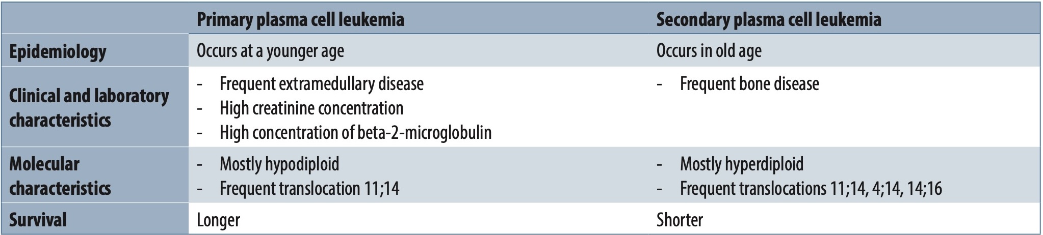
Plasma cell leukemia can be primary, occurring as a de novo disease (1%-2% of patients with multiple myeloma) and secondary, representing the leukemic transformation of refractory multiple myeloma or occurring in relapsed multiple myeloma (incidence 2%-9%) [6]. Regardless of whether it is primary or secondary plasma cell leukemia, the prognosis is poor. The biology and clinical manifestations of primary and secondary plasma cell leukemia differ (Table 1). Namely, patients with primary plasma cell leukemia show a better therapeutic response if they are treated with aggressive chemotherapy and autologous hematopoietic stem cell transplantation. However, unfortunately, relapses of the disease, which are most often refractory to therapy, are also registered within a short space of time [4]. On the other hand, secondary plasma cell leukemia shows a significantly worse therapeutic response (less than 50% of patients) with a swift fatal outcome [1]. Due to the introduction of new therapeutic modalities and the increase in the average life expectancy of patients suffering from multiple myeloma, the frequency of secondary plasma cell leukemia is increasing [4]. This increase in the frequency of secondary plasma cell leukemia can be explained by the higher prevalence of patients with multiple myeloma, which is a consequence of the extension of life expectancy, as well as the use of more therapeutic lines during the treatment of multiple myeloma, which can contribute to clonal selection and new genetic changes [7]. Nevertheless, primary plasma cell leukemia is the more common form of the disease and accounts for about 60%–70% of patients [2].
Biology of plasma cell leukemia
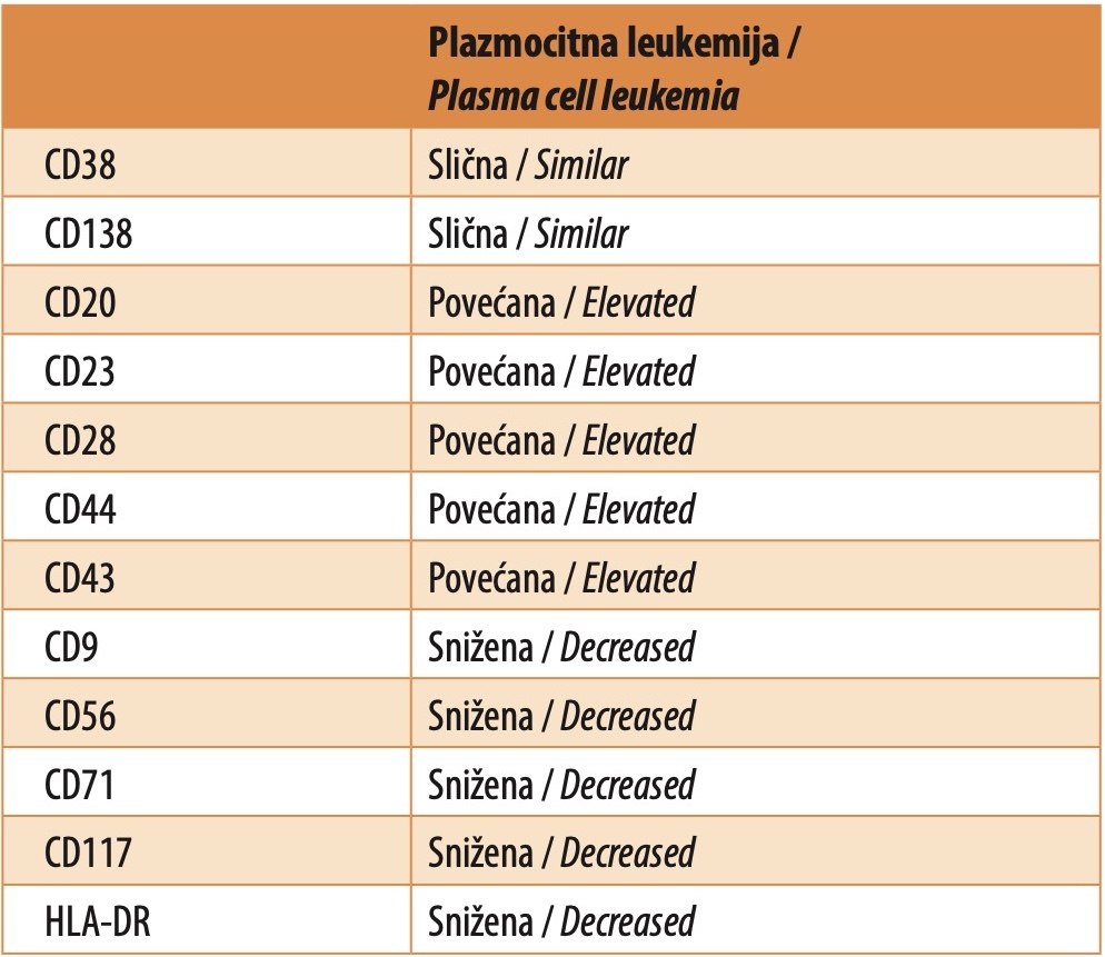
One of the main characteristics of most plasma cell dyscrasias is the proliferation of malignant plasma cells mainly into the bone marrow. Due to adhesion molecules expressed in plasma cells and cellular interactions with the bone marrow microenvironment, these malignant plasma cells most often remain in the bone marrow. In plasma cell leukemia, due to the altered expression of certain adhesion molecules, malignant plasma cells easily migrate from the bone marrow, enter the peripheral blood, and inhabit parenchymatous organs [8]. Plasmacytic leukemia cells show a reduced expression of NCAM molecules (neural cell adhesion molecule CD56), as well as of LFA (leukocyte function associated antigen-1), VLA-5 (very late antigen 5), CD9, CD82, and other adhesion molecules, thereby reducing the binding of plasmacytic leukemia cells to bone marrow stromal cells and consequently facilitating migration into the peripheral blood [9],[10].
The presence of numerous cytogenetic aberrations is typical for plasmacytic leukemia cells. Namely, plasmacytic leukemia cells are most often diploid or hypoploid (about 80%), while plasmacytes in multiple myeloma are most often hyperdiploid [3],[5]. The most common cytogenetic aberrations that occur in plasmacytic leukemia cells are deletion of chromosome 13 and monosomy which are found in more than 85% of patients [5],[10]. Chromosome 1 abnormalities (deletion or amplification) are also common in patients with plasma cell leukemia, especially in those with the primary form of the disease [5],[11],[12]. Translocation t(14;16) and deletion 17p are found in the majority of plasmacytic leukemia cells, which explains the frequent translocation of IgH as well as the expression of cyclin D1 [4]. Translocations t(11;14) and t(4;14) occur with approximately the same frequency as in patients with multiple myeloma [4]. Using the most modern molecular testing methods, a high frequency of TP53 and K/NRAS gene mutations has been determined [5]. The significance of each of these detected cytogenetic abnormalities is still insufficiently known, although they are believed to be associated with a more aggressive clinical course of the disease and a poorer prognosis.
Clinical manifestations
The clinical presentation of patients with plasma cell leukemia differs to some extent from the one found in multiple myeloma. The average age of patients with plasma cell leukemia is 61 years, which is about 10 years less than the average age of patients with multiple myeloma [4]. Most patients with plasma cell leukemia have a large tumor mass that causes weight loss, occasional night sweats, symptoms of kidney failure, anemia, and hemorrhagic syndrome. Namely, due to the large tumor mass and most often massive infiltration of the bone marrow, patients have severe anemia and thrombocytopenia. In addition, there is very often infiltration of peripheral organs, resulting in hepatosplenomegaly and lymphadenopathy, as well as infiltration of the lungs, pleura, testicles, central nervous system, and muscles with the presence of symptoms and clinical signs typical of infiltrated tissues [3],[4],[5],[6],[8],[9],[10]. Bone pain and osteolytic lesions occur somewhat less often than in patients with multiple myeloma [3],[4],[5],[6],[8],[9],[10].
Laboratory findings
Due to bone marrow infiltration, the largest number of patients with plasmacytic leukemia show the existence of normocytic anemia (about 80%) and thrombocytopenia (about 60%), and there is often leukocytosis with absolute plasmacytosis (above 2 x 10⁹ /L) [4],[6]. In the majority of patients, the rouleaux phenomenon is present in the peripheral blood smear [4],[6].
Biochemical analyses most often register increased serum activity of lactate dehydrogenase, and beta-2-microglobulin, as well as hypercalcemia, and an increase in serum creatinine and urea concentrations [6],[7]. In most patients, the presence of the M-component in the serum is registered, most often of the IgG class (about 50%), as well as IgA (about 15%), while IgD and IgE are very rare [6]. About 80% of patients with plasma cell leukemia have Bence-Jones proteinuria [6].
Bone marrow biopsy usually shows massive infiltration by plasmacytic leukemia cells with suppression of other hematopoietic lineages. In some patients, the morphology of plasmocytes is not changed, but in others, immature forms dominate, with a distinctly basophilic cytoplasm, two nuclei, a large nucleolus, and a perinuclear halo [1],[6],[8].
Diagnosis
The diagnosis of the disease is established using flow cytometry of peripheral blood. Namely, immunophenotypically, plasmacytic leukemia cells are CD38 and CD138 positive as is the case in multiple myeloma, but compared to plasma cells in multiple myeloma, they show a higher expression of CD20, CD23, CD28, CD44, and CD43 molecules, with reduced expression of CD9, CD56, CD71, CD117 and HLA-DR molecules (Table 2) [5],[8]. By detecting at least 5% of plasma cells with the above-mentioned immunophenotype among peripheral blood leukocytes, the diagnosis of plasma cell leukemia is established. After the diagnosis is made, it is necessary to take radiography of the skeleton or apply other radiological methods, such as computerized tomography (CT), nuclear magnetic resonance imaging (NMRI), or positron emission tomography (PET) of the whole body, to detect extramedullary disease. It is also necessary to determine, using fluorescence in situ hybridization (FISH), the existence of cytogenetic aberrations with particular reference to the presence of the deletion of chromosome 17p, deletion of chromosome 13q, deletion of chromosome 1p, and translocation of chromosome 14 (t(11;14), t(4;14), t(14;16)) [8].
Prognostic factors
Poor prognostic factors for patients with plasma cell leukemia are as follows: age over 60 years, thrombocytopenia below 100 x 109 /L, plasma cell count in peripheral blood greater than 20 x 109 /L, low serum albumin concentration, hypercalcemia, increased concentration of lactate dehydrogenase and beta-2-microglobulin, poorer performance status, as well as lack of therapeutic response to induction therapy [10],[12],[13],[14],[15]. In addition, the presence of hypodiploidy, complex karyotype, 13q deletion, 17p deletion, 1p deletion, and 1q amplification is associated with worse overall survival [11],[12]. Before the introduction of new agents and autologous transplantation, the average survival of patients with plasma cell leukemia was 4-11 months [9]. The introduction of new therapeutic modalities, as well as hematopoietic stem cell transplantation, has extended the average survival of patients and it is now 2-3 years [16].
Treatment
Bearing in mind the aggressiveness of the disease, it is necessary to start treatment immediately after diagnosis. There are certain differences in the treatment of primary and secondary plasma cell leukemia. The choice of therapeutic regimen mainly depends on whether the patient is a candidate for autologous transplantation or not.
The application of conventional chemotherapy in the treatment of primary plasma cell leukemia did not give satisfactory results, therefore, the induction therapy for plasma cell leukemia, regardless of whether the patient is a candidate for autologous transplantation or not, is based on the use of proteasome inhibitors and/or immunomodulatory drugs [15],[17]. Therapeutic regimens based on bortezomib in the treatment of primary plasma cell leukemia result in an overall response rate (ORR) of 76%–80% [2],[17]. The application of lenalidomide in combination with dexamethasone has also given encouraging results in treatment [16]. Even better results have been recorded with a combination of bortezomib, lenalidomide, and dexamethasone with the addition of elotuzumab [7]. The introduction of carfilzomib (second-generation proteasome inhibitors), in combination with lenalidomide and dexamethasone, leads to an overall therapeutic response in 93% of patients, with complete remission registered in 33% of patients [18].
If the patient is a candidate for autologous stem cell transplantation, after the application of induction high-dose chemotherapy, high-dose melphalan is administered as a conditioning regimen, and autologous transplantation is performed. With this therapy, complete remission is achieved in up to 40% of patients [19]. For younger patients with a matched donor, it is possible to continue treatment with allogeneic hematopoietic stem cell transplantation [20],[21]. After the completion of induction therapy and/or transplantation of hematopoietic stem cells, it is recommended to apply maintenance therapy using lenalidomide, bortezomib, or dexamethasone until disease progression [8].
The application of induction therapy carries the risk of tumor lysis syndrome. A large tumor mass and high proliferative potential of malignant plasma cells are risk factors for this treatment complication. Therefore, it is recommended that in this treatment phase, appropriate rehydration and uricosuppressors should be administered with continuous monitoring of the patient [9]. In addition, due to the use of proteasome inhibitors and immunomodulatory drugs, the use of prophylactic antiviral and anticoagulant therapy is advised [9].
Primary plasma cell leukemia relapse treatment depends on the patient’s age, performance status, therapeutic response to previous treatment, previously administered treatments, comorbidities, and renal insufficiency. Application of a combination of new therapeutic agents is definitely advised, but the therapeutic success is very low [8],[18],[21]. Literature data on the therapeutic success of modalities based on the use of bortezomib, carfilzomib, and monoclonal antibodies are very scarce. The treatment of secondary plasma cell leukemia depends on the patient’s age, comorbidities, previous therapeutic response, and the duration of the therapeutic response achieved with the previous therapy, therefore, there is still no gold-standard of treatment. Regardless of the applied therapeutic regimen, success is very low, and survival is usually only a few months [4].
Bearing in mind the very limited results in the treatment of primary and secondary plasma cell leukemia, it is expected that the application of new agents such as venetoclax, pomalidomide, ixazomib, selinexor, carfilzomib, daratumumab, anti-CD38 monoclonal antibodies, BRAF/MEK inhibitors, CAR-T therapy and other agents, could lead to an improvement in therapeutic response [4],[9]. The results from the studies published so far are very limited, but promising.
Informations

-
Keywords:
-
Received:
-
Revised:
-
Accepted:
-
Online first:
-
DOI:
-
Cite this article:

Predrag Đurđević
Clinic for Hematology, University Clinical Center Kragujevac
30 Zmaj Jovina Street, 34000 Kragujevac, Serbia
E-mail:
1. Jelinek T, Kryukov F, Rihova L, Hajek R. Plasma cell leukemia: from biology to treatment. Eur J Haematol. 2015 Jul;95(1):16-26. doi: 10.1111/ejh.12533. [CROSSREF]
2. Jung SH, Lee JJ. Update on primary plasma cell leukemia. Blood Res. 2022 Apr 30;57(S1):62-6. doi: 10.5045/br.2022.2022033. [CROSSREF]
3. Fernandez de Larrea C, Kyle RA, Durie BG, Ludwig H, Usmani S, Vesole DH, et al.; International Myeloma Working Group. Plasma cell leukemia: consensus statement on diagnostic requirements, response criteria and treatment recommendations by the International Myeloma Working Group. Leukemia. 2013 Apr;27(4):780-91. doi: 10.1038/leu.2012.336. [CROSSREF]
4. Gundesen MT, Lund T, Moeller HEH, Abildgaard N. Plasma cell leukemia: definition, presentation, and treatment. Curr Oncol Rep. 2019 Jan 28;21(1):8. doi: 10.1007/s11912-019-0754-x. [CROSSREF]
5. Rojas EA, Gutiérrez NC. Genomics of plasma cell leukemia. Cancers (Basel). 2022 Mar 21;14(6):1594. doi: 10.3390/cancers14061594. [CROSSREF]
6. Swami VK, Dwyer-Joyce LEA. Clinical pathology rounds: diagnosing plasma cell leukemia. Lab Med. 2000 Jun 1;31(6):312-5. doi: 10.1309/K6WB-G6GE82A9-KK4P. [CROSSREF]
7. Gowin K, Skerget S, Keats JJ, Mikhael J, Cowan AJ. Plasma cell leukemia: a review of the molecular classification, diagnosis, and evidenced-based treatment. Leuk Res. 2021 Dec;111:106687. doi: 10.1016/j.leukres.2021.106687. [CROSSREF]
8. van de Donk NW, Lokhorst HM, Anderson KC, Richardson PG. How I treat plasma cell leukemia. Blood. 2012 Sep 20;120(12):2376-89. doi: 10.1182/blood-2012-05-408682. [CROSSREF]
9. Tuazon SA, Holmberg LA, Nadeem O, Richardson PG. A clinical perspective on plasma cell leukemia: current status and future directions. Blood Cancer J. 2021 Feb 4;11:23. doi: 10.1038/s41408-021-00414-6. [CROSSREF]
10. Tiedemann RE, Gonzalez-Paz N, Kyle RA, Santana-Davila R, Price-Troska T, Van Wier SA, et al. Genetic aberrations and survival in plasma cell leukemia. Leukemia. 2008 May;22(5):1044-52. doi: 10.1038/leu.2008.4. [CROSSREF]
11. Singh ZN, Owens RB, Nair B, Shaughnessy JJr, Barlogie B. Loss of chemokine receptor expression and tetrasparins is correlated with extramedullary disease in multiple myeloma. Blood. 2010 Nov 19;116(21):2983. doi: 10.1182/ blood.V116.21.2983.2983. [CROSSREF]
12. Pagano L, Valentini CG, De Stefano V, Venditti A, Visani G, Petrucci MT, et al.; for GIMEMA-ALWP (Gruppo Italiano Malattie EMatologiche dell’Adulto, Acute Leukemia Working Party: coordinator Sergio Amadori). Primary plasma cell leukemia: a retrospective multicenter study of 73 patients. Ann Oncol. 2011 Jul;22(7):1628-35. doi: 10.1093/annonc/mdq646. [CROSSREF]
13. Yu T, Xu Y, An G, Tai YT, Ho M, Li Z, et al. Primary plasma cell leukemia: real-world retrospective study of 46 patients from a single-center study in China. Clin Lymphoma Myeloma Leuk. 2020 Oct;20(10):e652-9. doi: 10.1016/j.clml.2020.05.014. [CROSSREF]
14. Schinke C, Boyle EM, Ashby C, Wang Y, Lyzoguboiv V, Wardell C, et al. Genomic analysis of primary plasma cell leukemia reveals complex structural alterations and high-risk mutational patterns. Blood Cancer J. 2020 Jun 19;10(6):70. doi: 10.1038/s41408-020-0336-z. [CROSSREF]
15. Čolović M, Janković G, Suvajdžić N, Milić N, Đorđević V, Janković S. Thirty patients with primary plasma cell leukemia: a single center experience. Med Oncol. 2008;25(2):154-60. doi: 10.1007/s12032-007-9011-5. [CROSSREF]
16. Musto P. Progress in the treatment of primary plasma cell leukemia. J Clin Oncol. 2016 Jun 20;34(18):2082-4. doi: 10.1200/JCO.2016.66.6115. [CROSSREF]
17. Katodritou E, Terpos E, Delimpasi S, Kotsopoulou M, Michalis E, Vadikolia C, et al. Real-world data on prognosis and outcome of prim,ary plasma cell leukemia in the era of novel agents: a multicenter national study by the Greek Myeloma Study Group. Blood Cancer J. 2018 Mar 9;8(3):31. doi: 10.1038/ s41408-018-0059-6. [CROSSREF]
18. van de Donk NWCJ, Minnema MC, van der Holt B, Schjesvold F, Wu KL, Broijl A, et al. Treatment of primary plasma cell leukaemia with carfilzomib and lenalidomide-based therapy (EMN12/HOVON-129): final analysis of a non-randomised, multicentre, phase 2 study. Lancet Oncol. 2023 Oct;24(10):1119-33. doi: 10.1016/S1470-2045(23)00405-9. [CROSSREF]
19. Drake MB, Iacobelli S, van Biezen A, Morris C, Apperley JF, Niederwieser D, et al. Primary plasma cell leukemia and autologous stem cell transplantation Haematologica. 2010 May;95(5):804-9. doi: 10.3324/haematol.2009.013334. [CROSSREF]
20. Jung SH, Lee JJ, Kim K, Suh C, Yoon DH, Min CK, et al.; Korean Multiple Myeloma Working Party. The role of frontline autologous stem cell transplantation for primary plasma cell leukemia: a retrospective multicenter study (KMM160). Oncotarget. 2017 Jun 16;8(45):79517-26. doi: 10.18632/oncotarget.18535. [CROSSREF]
21. Mahindra A, Kalaycio ME, Vala-Ojeda J, Vesole DH, Zhang MJ, Li P, et al. Haematoietic cell transplantation for primary plasma cell leukemia: results from the Center for International Blood and Marrow Transplant Research. Leukemia. 2012 May;26(5):1091-7. doi: 10.1038/leu.2011.312. [CROSSREF]