


Lower eyelid dirofilariosis: A rare case of ocular parasitic infection with possible endemic exposure
- Eye Hospital, University Clinical Center of Serbia, Belgrade, Serbia
- University of Belgrade, Faculty of Medicine, Belgrade, Serbia
ABSTRACT
Introduction: Dirofilariosis, caused by the parasitic nematode Dirofilaria repens, is a zoonotic disease with rare ocular manifestations that are being increasingly reported in regions experiencing expanded parasite distribution. Infection in humans manifests as a localized inflammatory reaction and ocular involvement can lead to significant discomfort and diagnostic challenges. While often associated with travel to endemic areas, such as the Mediterranean region, dirofilariosis is also endemic in Serbia, which highlights the possibility of local transmission.
Case report: A 38-year-old female patient presented with unusual sensations of movement in her lower eyelid following a vacation in Greece. Clinical examination revealed localized swelling and a palpable moving mass in the lower eyelid. Surgical excision of the lesion was performed, and parasitological analysis confirmed the presence of Dirofilaria spp. Given that dirofilariosis is also endemic in Serbia, the possibility of pre-existing local infection before the patient’s trip cannot be excluded. The patient’s symptoms resolved completely after surgery, with no recurrence or complications noted during follow-up. This case underscores the need for considering parasitic infections in the differential diagnosis of eyelid masses, especially in patients who have recently traveled to or live in endemic areas.
Conclusion: This case highlights the clinical presentation and successful management of lower eyelid dirofilariosis, a rare ocular manifestation of Dirofilaria spp. infection. While a travel history to endemic regions can raise suspicion, endemicity in regions like Serbia necessitates factoring in the possibility of local transmission. Early detection and prompt surgical intervention are often curative. Further research is needed to understand local transmission dynamics and the ocular involvement of dirofilariosis in endemic areas, as well as complications that may arise due to inadequate diagnosis and treatment.
INTRODUCTION
Dirofilariosis is a zoonotic parasitic infection caused by nematodes belonging to the Dirofilaria genus, which primarily infect dogs and other carnivores. Human infections occur incidentally when infected mosquitoes transfer larvae to humans through bites. The two species most frequently implicated in human dirofilariosis are Dirofilaria immitis and Dirofilaria repens [1]. Dirofilaria immitis commonly causes pulmonary dirofilariosis, affecting the lungs and pulmonary arteries, whereas Dirofilaria repens is more frequently associated with subcutaneous and ocular infections [2].
In regions such as the Mediterranean basin, particularly in countries like Greece, Dirofilaria repens infections are facilitated by high mosquito prevalence and the presence of infected animals, i.e., reservoirs [3]. Serbia is also recognized as an endemic area for Dirofilaria repens, with numerous studies highlighting significant infections in both dogs and humans. Since 2008, systematic research has confirmed that the climate in Serbia, mosquito populations, and canine reservoirs create favorable conditions for endemic transmission. Tasić et al. documented canine dirofilariosis in northern Serbia [4], while Tasić-Otašević et al. and Momčilović et al. reported multiple cases of autochthonous human infections, including subcutaneous and subconjunctival forms [5],[6].
Though ocular dirofilariosis is uncommon, it presents a unique diagnostic challenge due to its variable clinical manifestations, which can include swelling, redness, discomfort, or sensations akin to a foreign body in the affected area [7]. In some instances, patients report a distinctive sensation of “something moving” beneath the skin or around the eye, attributed to the nematode’s motility within subcutaneous tissue [8].
In this instance, we report the case of a 38-year-old woman who presented with sensations of movement in her lower eyelid following her vacation on a Greek island. While her travel history initially raised suspicion of an imported case, the infection could also have been acquired in Serbia, where Dirofilaria repens is endemic. Considering the life cycle and growth rate of Dirofilaria repens, the possibility of autochthonous infection in Serbia cannot be excluded, which underscores the diagnostic complexity of such cases.
Despite the concerning nature of the symptoms, ocular dirofilariosis generally has a favorable prognosis following surgical removal of the parasite. Recurrence is rare, and patients typically achieve full recovery. However, with factors such as increased global travel, climate change, and expanding mosquito populations, the incidence of human dirofilariosis has been on the rise [8]. Early recognition of the clinical signs is crucial to avoid misdiagnosis and unnecessary procedures, enabling prompt removal of the parasite and resolution of symptoms.
This case emphasizes the importance of considering parasitic infections in the differential diagnosis of unexplained eyelid masses or movement sensations. Clinicians must remain vigilant not only in patients with a recent history of traveling to endemic regions but also in patients residing in or traveling to areas with established local vector populations, such as Serbia, where incidental human infections are being increasingly reported. The uncertainty as to whether this patient’s infection was imported or autochthonous further emphasizes the need for enhanced diagnostic awareness in both endemic and non-endemic settings.
CASE REPORT
A 38-year-old woman presented at our ophthalmology clinic complaining of an unusual sensation in the outer part of her lower eyelid. She reported that the sensation had started seven days after returning from a vacation spent on a Greek island. She had been on the island of Thasos from July 1 to July 18, and she first noticed the sensation in her eyelid on July 31. She described it as intermittent and stated that she felt as though “something was moving” inside the eyelid. The patient denied any pain, itching, or redness, and there were no associated systemic symptoms such as fever, fatigue, or lymphadenopathy. Her medical history was unremarkable, with no previous history of allergies, systemic diseases, or ocular surgeries. She had not been taking any medications, and she did not remember any mosquito bites during her holiday. The patient had not traveled outside Belgrade in the eight months before her holiday and reported no prior trips to the countryside, being near rivers, or being exposed to animals. She also did not recall any mosquito bites before this event.
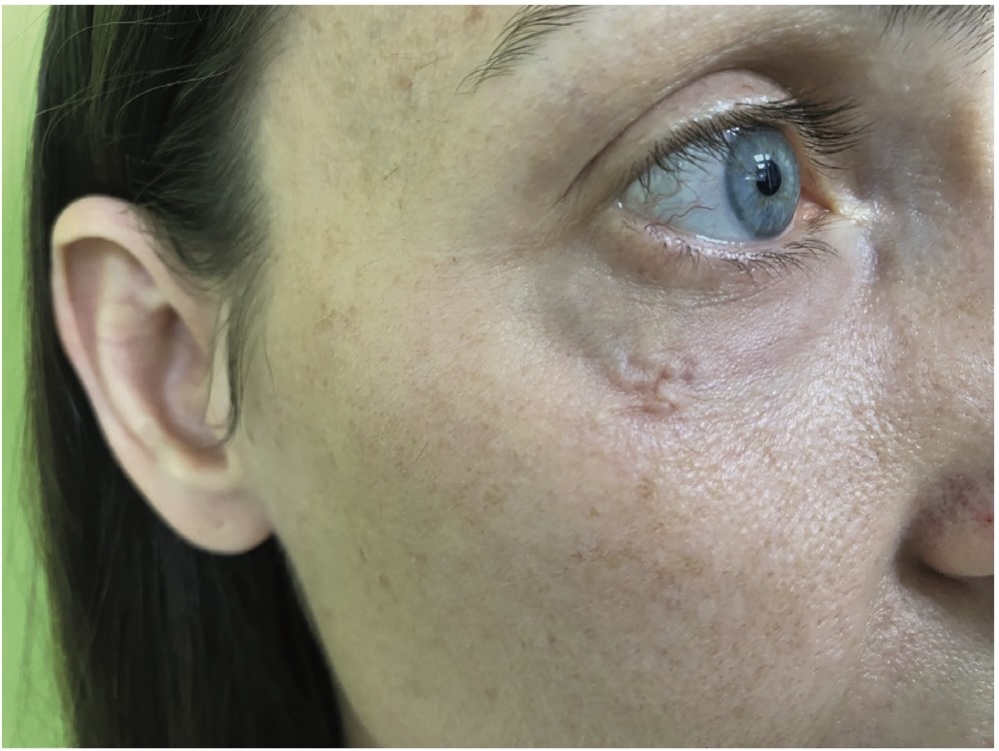
On examination, the patient’s visual acuity was 20/20 in both eyes. The anterior and posterior segments of the eyes were completely normal. There was no evidence of conjunctival hyperemia, discharge, or corneal abnormalities. Initial external inspection of the eyelid did not reveal any visible swelling, erythema, or abnormal masses. However, as the patient continued to describe the sensation of movement, a small, localized elevation became noticeable in the medial part of the lower eyelid. On closer observation, a worm-like structure was seen moving beneath the skin. Upon palpation, the structure disappeared but reappeared several minutes later in the temporal part of the lower eyelid (Figure 1).
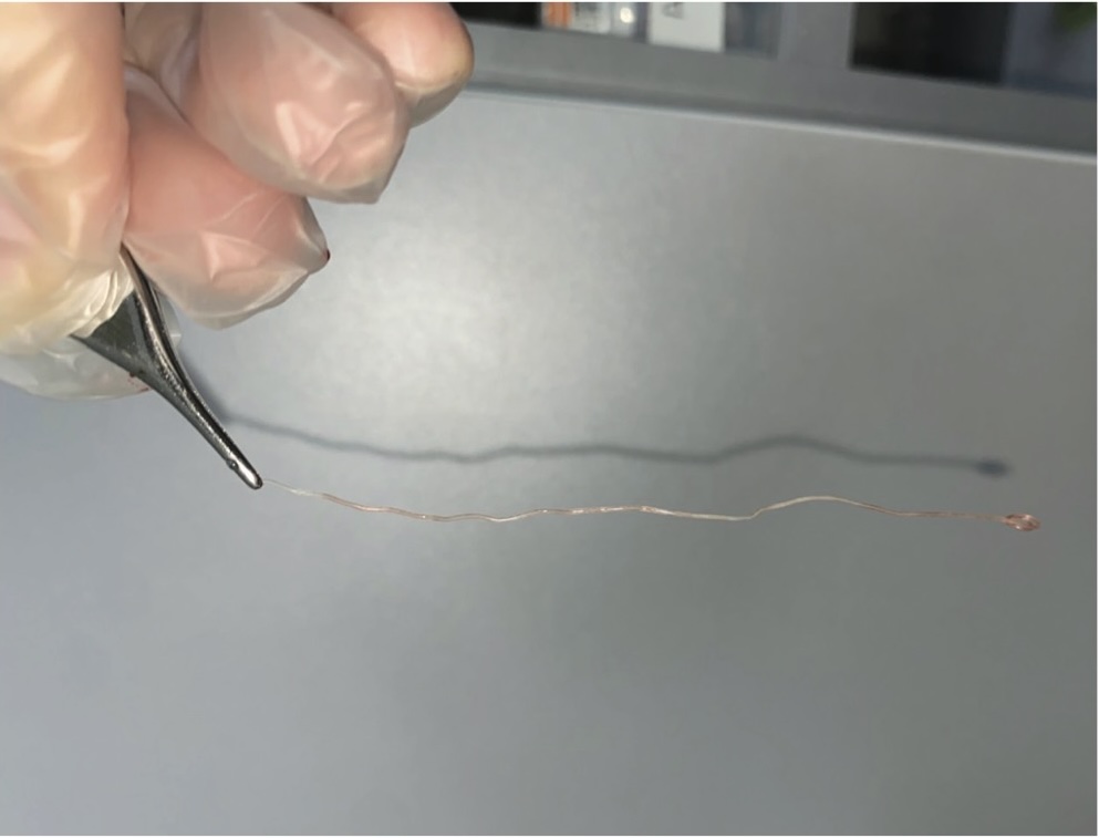
Given the clinical presentation, we suspected a parasitic infection. An infectious diseases specialist conducted initial blood tests, which showed no abnormalities, and the patient’s results were negative for eosinophilia. We informed the patient about the likely diagnosis and recommended immediate removal of the parasite. The patient consented to the procedure. Without the need for local anesthesia, we waited for the parasite to reappear under the skin. Once the movement was visible again, a small incision was made in the temporal part of the lower eyelid, and using fine forceps, the worm-like parasite was extracted. The nematode measured over 12 centimeters in length. The procedure was uncomplicated, and the patient did not experience any pain or discomfort during or after the removal (Figure 2). The extracted parasite was sent to the parasitology department for identification and the result confirmed Dirofilaria spp.
Following the extraction, the infectious diseases specialist conducted additional tests, including radiography of the heart and lungs, which showed normal results. Knott’s concentration method was also used to test for microfilariae in the blood, and the results were negative. IgE levels were within the normal range (below 4.7 IU/ml; reference range 0–100 IU/ml). The blood tests were repeated seven days after the initial test, with no significant changes observed. Given the absence of microfilariae and normal lab findings, additional antiparasitic treatment was not indicated and the infection was classified as an amicrofilaremic case of Dirofilaria spp. infection successfully treated surgically.
Postoperatively, the patient did not require any additional treatment, and the small incision healed well, leaving a barely visible scar. One month later, the patient’s clinical examination findings remained normal, with no signs of recurrence or additional symptoms.
DISCUSSION
Human infection with Dirofilaria occurs when a mosquito introduces an infective third-stage larva (L3) into the bloodstream when biting the host. While humans are accidental hosts and the larva rarely develops into an adult worm, in some cases, this progression is possible. In the patient we have described, the larva matured into its adult form, illustrating one of the uncommon scenarios in which this can happen.
Ocular dirofilariosis, though rare, must be taken into consideration as a differential diagnosis in patients presenting with unexplained eyelid swelling or sensations of movement, particularly in those who have traveled to regions endemic to Dirofilaria. In our case, the patient had recently visited a Greek island, which is part of the Mediterranean region known for endemic mosquito-borne infections. However, given the life cycle and growth rate of Dirofilaria repens, this infection could have been also acquired in Serbia, where autochthonous dirofilariosis is well documented [9]. The full life cycle of Dirofilaria repens, including larval stages and the prepatent period before microfilariae excretion, typically spans 6–9 months. In humans, Dirofilaria repens often fails to reach full maturity, yet the parasite’s twelve-centimeter length suggests that infection may have occurred several months earlier, making recent travel an unlikely origin.
In Serbia, dirofilariosis cases have been documented in both humans and animals, particularly in areas along river courses where mosquito vectors are prevalent. Džamić et al. reported cases of subcutaneous Dirofilaria repens infection in Serbia and Montenegro, including periocular sites, and highlighted the role of regional vectors in transmission [10]. The case of our patient, who had no recent exposure to the countryside and lived in an urban setting, demonstrates that urban cases are possible and that the urban setting should not exclude dirofilariosis as a differential diagnosis. Tasić et al. also noted significant canine infections in Serbia, underscoring the importance of zoonotic reservoirs in human exposure to Dirofilaria repens [11]. Given these findings and the patient’s limited travel history outside of Belgrade, endemic origin in Serbia remains plausible.
The pathogenesis of human dirofilariosis involves mosquito-borne transmission of infective larvae, which migrate through subcutaneous tissues and mature into adult worms, reaching 10 to 15 centimeters in length. Simon et al. reported that Dirofilaria repens can lodge in various periocular structures, such as the eyelid, orbit, and subconjunctival tissues, causing symptoms of movement [12]. Džamić et al. documented several cases wherein patients experienced sensations of movement in subcutaneous sites, suggesting that this mobility is characteristic of Dirofilaria repens infections [9].
Epidemiologically, recent studies have documented a rise in the number of ocular dirofilariosis cases in Mediterranean regions, driven by climate change and the mobility of humans, both of which expand mosquito vector habitats. Pupić-Bakrač et al. discussed human microfilaremia cases, underlining the importance of vector control in preventing infections [13]. The infection control implication is clear as awareness of Dirofilaria infection risk extends even to urban, non-endemic areas, which is in keeping with our case of suspected autochthonous dirofilariosis.
Surgical excision remains the cornerstone of ocular dirofilariosis treatment, offering a safe and effective approach with minimal complications. Kalogeropoulos et al. emphasized that excision typically resolves symptoms without the need for additional therapy. In our case, the absence of microfilariae in blood tests and a normal IgE level confirmed an amicrofilaremic infection, supporting the sufficiency of surgical management [3]. Similarly, Tasić-Otašević et al. documented cases where excision alone was appropriate, with no need for systemic therapy, which coincides with our patient’s outcome, where no recurrence was observed post-removal [14]. It is important to note that, based on general consensus, antihelmintic therapy is not typically recommended for cases of superficial dirofilariosis.
Diagnosing ocular dirofilariosis can be challenging due to its variable and nonspecific presentation. Engelsberg and Bläckberg highlighted the need to include parasitic infections in the differential diagnosis of periocular masses, particularly in patients from endemic areas or those with recent travel history [7]. Misdiagnoses can lead to inappropriate treatments, such as antibiotics or steroids, which are ineffective for parasitic infections. Additionally, Radovanović Spurnić et al. emphasized the importance of timely detection and surgical removal, to prevent unnecessary and potentially harmful management [15]. Our case emphasizes the need to consider Dirofilaria as a differential diagnosis not only in endemic regions but also in non-traveling patients with periocular symptoms.
Finally, the case of our patient highlights the importance of public and professional awareness of dirofilariosis risks in both endemic and emerging regions. Diagnosing this condition requires more than clinical suspicion; it necessitates the expertise of microbiologists specializing in parasitology and the use of advanced diagnostic tools, including radiological imaging, immunodiagnostics, and molecular techniques. Savić et al. conducted a seroepidemiological study in the northern part of Serbia, documenting significant seroprevalence in humans and canines and emphasizing the role of environmental and ecological factors in transmission [16]. Educating travelers and residents about preventive measures, such as avoiding mosquito exposure, remains critical. Clinicians in Serbia and other regions experiencing the spread of dirofilariosis must maintain a high level of awareness and consider this parasitic infection in cases presenting with periocular symptoms.
CONCLUSION
The presented case of ocular dirofilariosis highlights the need for clinicians to maintain a high index of suspicion for parasitic infections in patients presenting with atypical periocular symptoms, especially those with recent travel history to endemic regions or residing in areas with local vector populations. In the case of our patient, successful removal of the Dirofilaria spp. led to complete symptom resolution and minimal scarring. With the increase in global travel, climate change, and expanding mosquito habitats, it is crucial to recognize the potential for dirofilariosis in both endemic and non-endemic regions. Early diagnosis and timely surgical intervention are essential to avoid unnecessary treatments and to ensure optimal patient outcomes.
Informations

-
Keywords:
-
Received:
-
Revised:
-
Accepted:
-
Online first:
-
DOI:
-
Cite this article:

Dolika D. Vasović
Eye Hospital, University Clinical Center of Serbia
2 Pasterova Street, 11129 Belgrade, Serbia
E-mail:
1. Ying Z, Upadhyay A, Wang J, Han Q, Liu Q. The prevalence of canine dirofilariasis in China: a systematic review and meta-analysis. Parasit Vectors. 2023 Jun 20;16(1):207. doi: 10.1186/s13071-023-05770-9. [CROSSREF]
2. Camacho M, Antonietti M, Sayegh Y, Colson JD, Kunkler AL, Clauss KD, et al. Ocular Dirofilariasis: A Clinicopathologic Case Series and Literature Review. Ocul Oncol Pathol. 2024 Apr;10(1):43-52. doi: 10.1159/000533340. [CROSSREF]
3. Kalogeropoulos CD, Stefaniotou MI, Gorgoli KE, Papadopoulou CV, Pappa CN, Paschidis CA. Ocular dirofilariasis: a case series of 8 patients. Middle East Afr J Ophthalmol. 2014 Oct-Dec;21(4):312-6. doi: 10.4103/0974-9233.142267. [CROSSREF]
4. Tasić A, Rossi L, Tasić S, Miladinović-Tasić N, Ilić T, Dimitrijević S. Survey of canine dirofilariasis in Vojvodina, Serbia. Parasitol Res. 2008 Nov;103(6):1297- 302. doi: 10.1007/s00436-008-1132-z. [CROSSREF]
5. Tasić-Otašević SA, Trenkić Božinović MS, Gabrielli SV, Genchi C. Canine and human Dirofilaria infections in the Balkan Peninsula. Vet Parasitol. 2015 Apr 30;209(3-4):151-6. doi: 10.1016/j.vetpar.2015.02.016. [CROSSREF]
6. Momčilović S, Gabrielli S, Đenić N, Živković N, Stevanović G, Krstić M, et al. New cases of human dirofilariosis on the Balkan Peninsula - “Masked intruders” uncovered by a surgeon. Parasitol Int. 2022 Feb;86:102482. doi: 10.1016/j.parint.2021.102482. [CROSSREF]
7. Engelsberg K, Bläckberg J. Dirofilaria repens infection of the eye. J Ophthalmic Inflamm Infect. 2022 Mar 24;12(1):13. doi: 10.1186/s12348-022-00290-6. [CROSSREF]
8. Capelli G, Genchi C, Baneth G, Bourdeau P, Brianti E, Cardoso L, et al. Recent advances on Dirofilaria repens in dogs and humans in Europe. Parasit Vectors. 2018 Dec 19;11(1):663. doi: 10.1186/s13071-018-3205-x. [CROSSREF]
9. Džamić AM, Colović IV, Arsić-Arsenijević VS, Stepanović S, Boricić I, Džamić Z, et al. Human Dirofilaria repens infection in Serbia. J Helminthol. 2009 Jun;83(2):129-37. doi: 10.1017/S0022149X09341346. [CROSSREF]
10. Džamić AM, Arsić-Arsenijević V, Radonjić I, Mitrović S, Marty P, Kranjcić-Zec IF. Subcutaneous Dirofilaria repens infection of the eyelid in Serbia and Montenegro. Parasite. 2004 Jun;11(2):239-40.
11. Tasić A, Tasić-Otašević S, Gabrielli S, Miladinović-Tasić N, Ignjatović A, Dorđević J, et al. Canine Dirofilaria infections in two uninvestigated areas of Serbia: epidemiological and genetic aspects. Vector Borne Zoonotic Dis. 2012 Dec;12(12):1031-5. doi: 10.1089/vbz.2011.0949. [CROSSREF]
12. Simon F, Siles-Lucas M, Morchon R, Gonzalez-Miguel J, Mellado L, Carreton E, et al. Human and animal dirofilariasis: the emergence of a zoonotic mosaic. Clin Microbiol Rev. 2012;25:507–544. doi: 10.1128/CMR.00012-12. [CROSSREF]
13. Pupić-Bakrač A, Pupić-Bakrač J, Beck A, Jurković D, Polkinghorne A, Beck R. Dirofilaria repens microfilaremia in humans: Case description and literature review. One Health. 2021 Aug 12;13:100306. doi: 10.1016/j.onehlt.2021.100306. [CROSSREF]
14. Tasić-Otašević S, Golubović M, Trichei S, Zdravković D, Jordan R, Gabrielli S. Microfilaremic Dirofilaria repens Infection in Patient from Serbia. Emerg Infect Dis. 2023 Dec;29(12):2548-2550. doi: 10.3201/eid2912.230796. [CROSSREF]
15. Radovanović Spurnić AP, Stevanović G, Dakić Z, Ofori Belić I, Spurnić I, Pelemiš M. Human subcutaneous dirofilariasis – case report. Med Pregl. 2015 JulAug;68(7-8):273-6. doi: 10.2298/mpns1508273r. [CROSSREF]
16. Savić S, Stosić MZ, Marčić D, Hernández I, Potkonjak A, Otašević S, et al. Seroepidemiological Study of Canine and Human Dirofilariasis in the Endemic Region of Northern Serbia. Front Vet Sci. 2020 Sep 29;7:571. doi: 10.3389/ fvets.2020.00571. [CROSSREF]
- Picture 1. Preoperative image of a 38-year-old female patient showing the right lower eyelid with visible slight swelling indicative of a subcutaneous parasite (Dirofilaria spp.). The patient experienced discomfort and the sensation of movement within the eyelid, correlating with parasitic presence, before surgical removal
- Picture 2. A thin, elongated parasitic organism (Dirofilaria spp.) is shown being held with tweezers after surgical extraction. The worm-like structure is 12 centimeters long