


Penoscrotal Paget's disease – Case report and literature review
- Institute of Oncology and Radiology of Serbia, Belgrade, Serbia
- University of Belgrade, Faculty of Medicine, Belgrade, Serbia
ABSTRACT
Introduction: Penoscrotal Paget’s disease is very rare worldwide, with a small number of case reports, and it thus represents a major dilemma in diagnosis and treatment.
Case report: An 81-year-old man presents himself to the Institute of Oncology and Radiology of Serbia (IORS) for the first time due to an ulcerated and erythematous tumor change about 10 cm in diameter covered with squamous plates, which extends from the groin region on the left to the scrotum. The tumefaction is erythematous, bloody and it occasionally bleeds, and the patient complains of occasional pain in the left leg. Previously, a biopsy of the mentioned tumor change was performed in another institution, and the PH finding was in favor of extramammary Paget’s disease. The patient was presented to the IORS council, which decided on operative treatment.
Conclusion: This rare disease is also found outside of clinically visible lesions, positive resection margins are common, and this represents the main risk factor for disease recurrence. Accurate preoperative diagnosis and proper surgical treatment are crucial for preventing relapses and repeat operations, making them key factors in improving survival. This case report certainly represents a significant step for oncologists in understanding this disease.
INTRODUCTION
Extramammary Paget’s disease (EMPD) is a rare intraepithelial malignancy that typically occurs in areas rich in apocrine glands, such as the perineum, vulva, axilla, scrotum, and penis [1]. The exact pathogenesis of EMPD is still not completely understood. Current data indicate that EMPD is a heterogenous disease that can be primary and secondary. Primary EMPD originates from the skin, more precisely from the epidermis or apocrine sweat glands. This form is initially limited to the epithelium but can progress to an invasive tumor that spreads to the dermis, as well as blood and lymphatic vessels. In advanced stages, it may lead to lymph node or visceral metastases. The secondary form of EMPD is associated with the spread of malignant adenocarcinoma cells originating from dermal adnexal glands or adjacent epithelium, typically from the genitourinary or gastrointestinal tract [2]. The incidence of extramammary Paget’s disease is 6 per 1,000,000 people per year [3], with penoscrotal EMPD accounting for only 14% of all EMPD cases [4].
We present the case of an 81-year-old man with penoscrotal EMPD.
CASE
An 81-year-old man presented to the Institute of Oncology and Radiology of Serbia (IORS) on 9th February 2023 with a first-time complaint of an ulcerated, erythematous tumor measuring approximately 10 cm in diameter, covered with squamous plaques and extending from the left groin to the scrotum. The mass is erythematous and bloody, with occasional bleeding, and the patient reports occasional pain in the left leg. A biopsy of the tumor was previously performed at another institution, with the histopathology results indicating extramammary Paget’s disease. The patient was presented to the IORS council, which recommended surgical treatment.
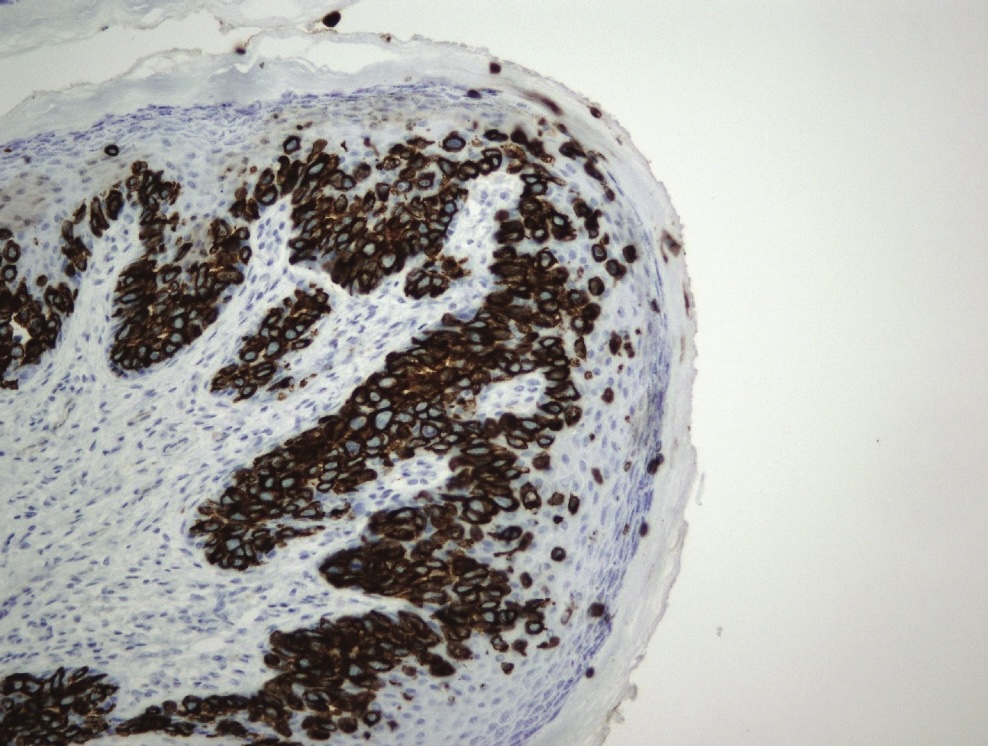
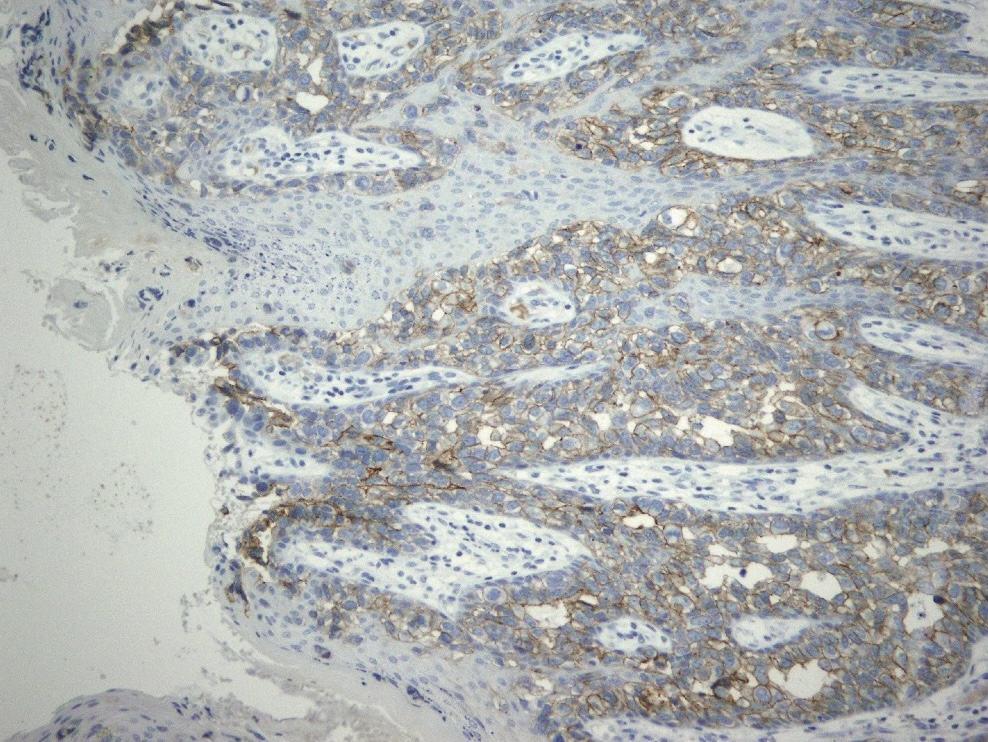
An excision of the tumor in the left inguinoscrotal region was performed, followed by primary closure of the defect. The histopathology report indicates the following immunohistochemical profile of the tumor cells: CK7+, HER2+, CK5/6+, HMB45-, p63-. The analyzed preparation reveals a tumor that, based on its histomorphological and immunohistochemical characteristics, is consistent with Paget’s disease. The tumor invades all layers of the dermis but does not extend into the subcutaneous fat tissue, and no lymphovascular invasion was observed. The lateral edges of the resection are infiltrated with tumor tissue, but the infiltration does not extend to the depth, classified as pT2.
The patient was regularly monitored postoperatively. After 4 months, he returned for a follow-up examination, during which an ultrasound of the inguinal region revealed two suspicious lesions, measuring 11 mm and 8 mm, with an indeterminate etiology. The patient was then referred to the council, which decided to proceed with a multidetector computed tomography (MDCT) scan.
The MDCT examination of the abdomen and pelvis revealed multiple focal lesions in the liver, consistent with secondary deposits. Lymphadenopathy was present retroperitoneally. An irregularly shaped prostate with extremely heterogeneous and indistinct contours, measuring 70 x 65 x 61 mm, was observed. It was compressing the urinary bladder and lacked a clearly defined fat plane adjacent to the right seminal vesicle. The bony structures of the lumbar spine showed rarefaction with osteolytic, patchy, and confluent areas. The L2 and L3 vertebral bodies were notably affected, exhibiting reduced height in the region of a pathological fracture. At that time, the patient reported bone pain in both the right and left groin, along with weakness in the lower extremities, which had persisted for 3 weeks.
The patient was presented again to the IORS council, which, after reviewing the MDCT findings, the patient’s reported symptoms, and his performance status (PS3), decided to conduct a comprehensive diagnostic work-up. This included PSA, CEA, and CA19-9 tests, a urological examination, colonoscopy, mammography, and MDCT of the thorax.
The mammographic findings were normal (BIRADS DD2 LD2). CEA tumor markers were as follows: CEA 435.38 ng/mL; CA19-9 59 U/mL; PSA 4.41 ng/mL. The MDCT examination of the thorax identified a subpleural nodule in the lung, measuring up to 7 mm, located from the mediastinal region. This finding, which could represent a secondary deposit or granuloma, warranted further follow-up. Due to the patient’s severe general condition (PS4), the urological examination and colonoscopy were not conducted.
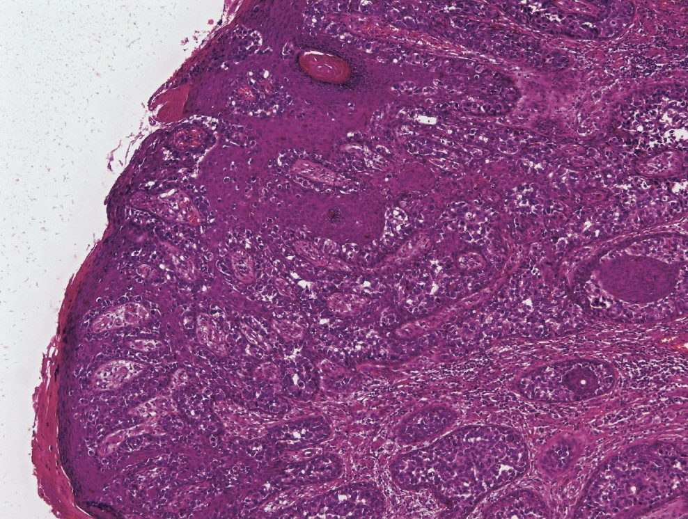
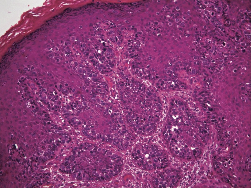
Figures 1-5 display histological sections of a tumor diagnosed with extramammary Paget’s disease.

The patient was reviewed by the council again, which, considering the patient’s overall condition and disease progression, concluded that specific oncological treatment was not indicated. Instead, they recommended continuing with symptomatic and supportive therapy.
Due to bone pain, pain in the left and right groin, and weakness in the lower extremities, the patient was evaluated at the IORS pain clinic. Symptomatic therapy was prescribed, including: 1. Durogesic patch 25 µg/h, 2. Oramorph syrup 0.2%, 3. Fresubin 2 kcal, 4. Fortecortin tablets 4 mg, 5. Lactulose syrup, and 6. Clometol tablets.
DISCUSSION
From a historical point of view, Crocker was the first to describe EMPD of the scrotum and penis in 1889 [5]. The clinical manifestation of this disease typically presents as a non-specific, well-demarcated erythematous plaque that may be thickened or covered with white, scaly patches, often with irregular borders [6],[7]. EMPD must be differentiated from other conditions, including squamous cell carcinoma, melanoma, and benign papulosquamous diseases. Penoscrotal lesions may initially develop in the groin and subsequently spread to the scrotum and penis. Lesions are most often accompanied by itching, but burning and swelling may also occur. In some cases, inguinal lymphadenopathy may be diagnosed, which can be associated with leg edema [8].
As mentioned in the introduction, EMPD is a rare condition with an incidence of only 6 per 1,000,000 people [3]. The vulva is the most common site for the disease, affecting approximately 65% of patients [9]. Compared to vulvar EMPD, penoscrotal EMPD occurs much less frequently. In the period between 1973 and 2002, only 100 cases were presented in the United States of America [10]. Macroscopically, the lesions present as well-circumscribed erythematous plaques that may itch, ulcerate, and bleed. Due to its clinical presentation, the condition is often misdiagnosed as eczema, lichen sclerosus, contact dermatitis, seborrheic dermatitis, fungal infection, or, most commonly, psoriasis. This frequent misdiagnosis can result in delays in reaching the correct diagnosis, referrals to inappropriate specialists, and incorrect treatments [4],[11]. If lesions in areas rich in apocrine glands do not respond to the previously prescribed therapy, EMPD should always be considered [12].
In a study conducted by scientists from Johns Hopkins University in Baltimore, the average age of patients with penoscrotal EMPD was 73 years, ranging from 63 to 87 [13].
A diagnosis of EMPD requires a histopathological examination of the biopsied tissue. The diagnosis is confirmed by the presence of Paget’s cells on routine hematoxylin-eosin staining. Most cases of EMPD are primary adenocarcinomas that probably originate from pluripotent cells in the basal layer of the epidermis. Unlike primary EMPB, secondary EMPB is associated with other anatomically close malignancies. The majority of malignancies associated with EMPD of the penis and scrotum are linked to genitourinary tract cancers, particularly prostate and bladder cancer, as well as rectal cancer [14].
The prognosis for localized EMPD is significantly better than for invasive disease, which is why the treatment approaches differ. For noninvasive EMPD, treatment options include surgical resection, radiotherapy, topical chemotherapy, and phototherapy. The most effective treatment for non-invasive EMPD is wide excision of the lesion, with most experts recommending margins of 1-3 cm in healthy tissue. Since EMPD often extends beyond macroscopically visible boundaries, Mohs micrographic surgery is recommended, as it offers a lower rate of local disease recurrence (23% vs 33%). Dissection of the associated lymph nodes is required if they are involved. Prophylactic lymph node dissection is not recommended, as it has no benefit.
In patients with invasive IMPB, surgery is not a curative treatment, so adjuvant chemotherapy and radiotherapy are often necessary. Cytotoxic agents such as 5-fluorouracil and Mitomycin C are effective for treating inadequately excised and advanced EMPD [15].
In a study conducted by an Asian research group involving 95 patients treated for EMPD between 1993 and 2020, the five-year survival rate was 79.3%, and the ten-year survival rate was 64.7%. Patients with secondary EMPD, due to the presence of an associated second malignancy, had a significantly shorter survival [16].
CONCLUSION
Extramammary Paget’s disease is a rare condition that affects skin areas rich in apocrine glands, including the vulva, scrotum, penis, and axilla. Because the disease often extends beyond clinically visible lesions, positive resection margins are common, which is a major risk factor for disease recurrence. Currently, the preferred therapies are wide local excision of the lesion or Mohs micrographic surgery. The rarity of this condition makes research focused on developing effective treatments particularly challenging. Larger studies are needed to provide more guidance on how to approach and treat patients with EMPD.
Informations

-
Keywords:
-
Received:
-
Revised:
-
Accepted:
-
Online first:
-
DOI:
-
Cite this article:

Luka Pejnović
Institute of Oncology and Radiology of Serbia
14 Pasterova Street, 11000 Belgrade, Serbia
E-mail:
1. Isrow D, Oregel KZ, Cortes J, Gomez H, Milikowski C, Feun L et al. Advanced Extramammary Paget's disease of the groin, penis, and scrotum. Clin Med Insights Oncol. 2014 Jul 6; 8:87-90. doi: 10.4137/CMO.S13107. [CROSSREF]
2. Kanitakis J. Mammary and extramammary Paget's disease. J Eur Acad Dermatol Venereol. 2007 May;21(5):581-90. doi: 10.1111/j.1468-3083.2007.02154.x. [CROSSREF]
3. Master VA, Herrel L, Johnson TV, Delman KA. Extramammary Paget's disease of the penis and anogenital area: SEER analysis. J Clin Oncol. 2011 Mar 1;29(7 Suppl): S220. doi:10.1200/jco.2011.29.7_suppl.220. [CROSSREF]
4. Ekwueme KC, Zakhour HD, Parr NJ. Extramammary Paget's disease of the penis: a case report and review of the literature. J Med Case Rep. 2009 Jan 6;3:4. doi: 10.1186/1752-1947-3-4. [CROSSREF]
5. Crocker HR. Paget’s disease affecting the scrotum and penis. Trans Pathol Soc Lond. 1889; 40:187-91.
6. O'Connor EA, Hettinger PC, Neuburg M, Dzwierzynski WW. Extramammary Paget's disease: a novel approach to treatment using a modification of peripheral Mohs micrographic surgery. Ann Plast Surg. 2012 Jun;68(6):616-20. doi: 10.1097/SAP.0b013e31821b6c7b. [CROSSREF]
7. Badgwell C, Rosen T. Treatment of limited extent extramammary Paget's disease with 5 percent imiquimod cream. Dermatol Online J. 2006 Jan 27;12(1):22. [CROSSREF]
8. Hoch WH. Adenocarcinoma of the scrotum (extramammary Paget's disease): case report and review of the literature. J Urol. 1984 Jul;132(1):137-9. doi: 10.1016/s0022-5347(17)49501-2. [CROSSREF]
9. Siesling S, Elferink MA, van Dijck JA, Pierie JP, Blokx WA. Epidemiology and treatment of extramammary Paget disease in the Netherlands. Eur J Surg Oncol. 2007 Oct;33(8):951-5. doi: 10.1016/j.ejso.2006.11.028. [CROSSREF]
10. Wright JL, Morgan TM, Lin DW. Primary scrotal cancer: disease characteristics and increasing incidence. Urology. 2008 Nov;72(5):1139-43. doi: 10.1016/j. urology.2008.06.043. [CROSSREF]
11. Li YC, Lu LY, Yang YT, Chang CC, Chen LM. Extramammary Paget's disease of the scrotum associated with hepatocellular carcinoma. J Chin Med Assoc. 2009 Oct;72(10):542-6. doi: 10.1016/S1726-4901(09)70425-3. [CROSSREF]
12. Heymann WR. Extramammary Paget's disease. Clin Dermatol. 1993 JanMar;11(1):83-7. doi: 10.1016/0738-081x(93)90101-h. [CROSSREF]
13. Shabihkhani M, Patil P, Murati Amador B, Plaza JA, Osunkoya AO, Lombardo KA et al. Extramammary Paget disease of the scrotum: a contemporary clinicopathologic analysis of 20 cases in the United States. Appl Immunohistochem Mol Morphol. 2020 Aug;28(7):524-31. doi: 10.1097/PAI.0000000000000789. [CROSSREF]
14. Payne WG, Wells KE. Extramammary Paget's disease of the scrotum. Ann Plast Surg. 1994 Dec;33(6):669-71. doi: 10.1097/00000637-199412000- 00021. [CROSSREF]
15. Juang GD, Lin MY, Hwang TI. Extramammary Paget's disease of the scrotum. J Chin Med Assoc. 2011 Jul;74(7):325-8. doi: 10.1016/j.jcma.2011.05.010. [CROSSREF]
16. Ha DL, Ha GU, Kim JY, Han MH, Lee HJ, Hong DG et al. J. Five- and 10-year survival in extramammary Paget's disease: a focus on wide local excision. Asia Pac J Clin Oncol. 2023 May 3. doi: 10.1111/ajco.13964. [CROSSREF]
- Picture 1. Extramammary Paget’s disease. Neoplastic cells with abundant cytoplasm are arranged in confluent nests and as single cells throughout the epidermis (H&E, 10x).
- Picture 2. Extramammary Paget’s disease. The clusters and single Paget cells compress the basal keratinocytes against the basement membrane (H&E, 20x).
- Picture 3. Extramammary Paget’s disease. Neoplastic cells show cytoplasmic and membranous CK7 staining (CK7, 20x).
- Picture 4. Extramammary Paget’s disease. Neoplastic cells show moderate, complete membranous staining for HER2 (HER 2 , 20x).
- Picture 5. Extramammary Paget’s disease. Neoplastic cells are negative for p63, whereas epidermal cells show nuclear p63 positivity (p63, 20x)