


Disease progression in patients with low-risk primary myelofibrosis – case report
- University Clinical Center of Serbia, Clinic for Hematology Belgrade, Serbia
- University of Belgrade, Faculty of Medicine, Belgrade, Serbia
ABSTRACT
Introduction: The median survival (OS) of patients with low-risk primary myelofibrosis (PMF) is over 15 years, but according to the “MOST” prospective study, disease progression occurs in almost 60% of these patients.
Aim: to present the disease course and treatment outcome of patients with lowrisk PMF. Case report: All patients were diagnosed with PMF, low-risk International Prognostic Scoring System (IPSS), and normal initial cytogenetics.
Case 1: A 61-year-old male patient was admitted in May 2016 with Tr 772x109 /L, LDH 566U/L, no splenomegaly, JAK2V617F+. He was initially treated with hydroxyurea (HU) from 2016 to 2020. In 2021, he was introduced to ruxolitinib due to the development of splenomegaly and leukocytosis, with disease progression 18 months later (spleen 26cm, Plt 20x109 /L, 10% blasts in the marrow, complex karyotype: -5, del 7q, mar+). Azacitidine was introduced, but death occurred due to sepsis in October 2022. OS is 66 months.
Case 2: A 47-year-old female patient was admitted in July 2011 with Tr 899x109 /L, LDH 899U/L, spleen 15x7cm, JAk2V617F+, and ASXL+. She was initially treated with HU and has been treated with ruxolitinib since October 2013 due to the progression of splenomegaly (spleen 19.3cm, LDH 1881U/L), with subsequent normalization of spleen size, number of Tr, and LDH. The patient is in remission. OS is 126 months.
Case 3: A 64-year-old male patient was admitted in May 2012 with Tr 1457x109 /L, LDH 631U/L, borderline splenomegaly, JAk2V617F-, MPL+, SRSF2+, U2AF1+, ASXL1+. He was initially treated with HU (May 2012) and since April 2019 with danazol due to the emergence of transfusion dependence. Due to heart failure, death occurred in February 2020. OS is 96 months.
Conclusion: Some patients with low-risk PMF have disease progression and future studies will show whether early Next Generation Sequencing (NGS) analysis of non-driver mutations and early initiation of therapy contribute to changing the course of the disease.
INTRODUCTION
Primary myelofibrosis (PMF) is an acquired clonal disorder of hematopoietic stem cells with abnormal proliferation of myeloid cells. PMF is a Bcr-Abl (Breakpoint cluster region protein-Abelson tyrosine-protein kinase, Eng.) negative myeloproliferative neoplasm (MPN) [1],[2]. It is characterized by the presence of constitutional symptoms, splenomegaly in the midst of extramedullary hematopoiesis, and the possibility of leukemic transformation [1]. Most patients have a Janus kinase gene mutation (JAK)2/V617F) [3]. The diagnosis of PMF is determined according to the ICC criteria (2022). The presence of nucleated erythrocytes, immature granulocytes, and dacryocytes is typical, but prefibrotic PMF does not necessarily show clear leukoerythroblastosis. Bone marrow fibrosis is usually associated with JAK2, CALR, or MPL mutations. The karyotype most often shows abnormalities of type +9, del(13k), del(20k), and chromosome number 1 [4],[5],[6]. The prognostic model is based on the development of the International Prognostic Scoring System (IPSS) from 2009, which is used in the initial diagnosis and includes age >65 years, hemoglobin 25x109 /l, presence > 1% of circulating blasts and development of constitutional symptoms. Depending on the number of harmful factors present, patients were stratified into groups of low, medium-1, medium-2, and high risk of the disease [7]. DIPSS (dynamic IPSS score) uses the same prognostic variables but is applicable at any time during the disease [8]. Conventional treatment has a limited impact on patient survival and includes “watch and wait” for asymptomatic patients, erythropoiesis stimulators, androgens or immunomodulators for anemia, the use of cytoreductive drugs (hydroxyurea in the development of splenomegaly and constitutional symptoms), and in individual cases, splenectomy or radiotherapy [9]. Targeted molecular therapy (JAK inhibitors) is effective in both JAK2+ and JAK2- patients. Ruxolitinib is the therapy of choice for PMF with splenomegaly and/or the development of constitutional symptoms. Allogeneic stem cell transplantation remains the only cure for PMF but is limited to high- and intermediate-2-risk patients. Many studies of the combination of JAK inhibitors with other agents are underway [9]. Several studies have shown that treatment of myelofibrosis earlier in the course of the disease, while the patient is at low or intermediate-1 risk, or as early as possible if he has intermediate and high risk, results in greater relative efficacy and less toxicity compared to later initiation of treatment [10]. This work aimed to present patients with low-risk PMF, in whom biological progression of the disease occurred, and to highlight their clinical and biological characteristics.
CASE REPORTS
First case
In a patient, male, age 61, in May 2016, leukocytosis and thrombocytosis were found in the blood count (Hb 158 g/L, MCV 90fl, Le 12.9x10⁹ /L, Tr 792x109 /L, in the differential formula neutrophils 66%, lymphocytes 26%, without leukoerythroblastosis), and in biochemical analyzes elevated LDH (556U/L). Abdominal ultrasound did not show the presence of splenomegaly. The patient did not have any symtoms during the examination. In the performed hematological diagnosis, the findings of the bone marrow biopsy showed fibrosis gr I, spontaneous development of erythroid colony formations was observed, and conventional cytogenetics was normal (20 metaphases). JAK2V617F mutation was detected (PCR). The serum erythropoietin level was average. A diagnosis of prefibrotic PMF, low risk according to the IPSS score, with an MPN 10 score of 10/100, was made.
Along with the use of acetylsalicylic acid of 100 mg per body per day, after the implementation diagnosis and further progression of thrombocytosis and leukocytosis, treatment with hydroxyurea (HU), 500 mg per body per day, was started (June 2016), in the period from 2016 to 2020. In December 2019, the follow-up abdominal ultrasound was still normal. However, in August 2020 (53 months after the diagnosis of PMF), a progression of leukocytosis was noted (30x109 /L, with Hb 129 g/L and Tr 161x109 /l), and the dose of HU was escalated to 1000 mg per person per day. In October 2020 (55 months after the diagnosis of PMF), the patient reported nausea, weight loss of 5 kg in one month, night sweats, and sweating. Further progression of leukocytosis (50x109 /L, 3% blasts in peripheral smear) and LDH increase (1627 U/L) were constant. The dose of HU was escalated to 1500 mg per os per day, with the subsequent occurrence of thrombocytopenia.
In October 2020, a hematological reevaluation was carried out. Aspiration puncture showed no acute transformation of MPN (2% blasts). In the bone marrow biopsy, fibrosis grade II was observed in 4% CD34+ cells. The findings of conventional cytogenetics in 20 mitoses were normal. The presence of PMF, intermediate-2 risk, according to the DIPSS score, and the MPN score of 19/100 was established. In February 2021, further progression of splenomegaly (22.2 cm) was detected radiographically, without splenic thrombosis. Ruxolitinib® was introduced at a dose of 2x20 mg per body per day, and a month later, due to leukocytosis 146x109 /l (with Hb 120 g/L and Tr 101x109 /l), HU was added again (500 mg per os per day). After 18 months from the start of ruxolitinib administration (August 2022), further progression of the disease was noted (deepening thrombocytopenia, progression of splenomegaly up to 26 cm), with the appearance of an aberrant karyotype with 2 independent clones (-5, del 7q, +mar). A repeat marrow aspiration showed 10% blasts in the marrow. The patient started treatment with azacitidine, but due to the appearance of a septic condition with the development of septic shock, the patient died in October 2022. The overall survival (OS) period was 66 months.
Second case
A 47-year-old patient consulted a hematologist in July 2011 due to the appearance of pruritus and abdominal pain, as well as blood count findings (Hb 128 g/L, MCV 89fL, Le 8.7x10⁹ /L, Tr 772x109 /l, in the differential formula neutrophils 66%, lymphocytes 25%, monocytes 5%, without leukoerythroblastosis). In laboratory analyses, LDH was 899 U/L. The initial abdomen ultrasound showed a spleen with a diameter of 15x7cm. A hematological diagnosis was carried out. In the bone marrow biopsy, Gr I fibrosis was observed, there was a positive, spontaneous development of erythroid colony formations, and conventional cytogenetics were normal (metaphase in 4 analyzed - low mitotic index). JAK2V617F mutation-homozygote was established (PCR). NGS (Next Generation Sequencing - Prof. Skoda mutation panel) analysis determined the presence of ASXL1 mutation. The serum erythropoietin level was average. The diagnosis of prefibrotic PMF, low risk, according to the IPSS score, and MPN 10 score 25/100 was established.
Acetylsalicylic acid (100 mg per os per day) was initially introduced, and from July 2012, HU (500 mg per os per day) was introduced, considering progressive splenomegaly (18x7cm) and an increase in LDH (1221U/L). Then, the presence of intermediate-1 risk was determined according to the DIPSS score. In October 2013, the patient reported malaise, night sweats, sweating, and the recurrence of abdominal pain. The spleen was palpated up to the level of the umbilicus. In the blood count, Hb 130x109 /L, Le 13x109 /l, Tr 563x109 /L, there is a tendency for further growth of LDH (1881U/L), along with normal other inflammatory parameters. Control ultrasound of the abdomen showed further progression of splenomegaly (19.3x7.5 cm). The patient was not motivated to undergo a bone marrow rebiopsy. The presence of intermediate-1 risk (DIPSS) was still established, with an increase in the MPN 10 score to 36/100.
In the same month, ruxolitinib was introduced at a dose of 2x20 mg per os per day (within the “compassionate” program). In further treatment with ruxolitinib, there is a complete regression of splenomegaly (13x5 cm in February 2015), with complete laboratory parameters. At the last control, in January 2024 (still ruxolitinib 2x20 mg per os per day), in the blood count Hb 124g/L, Le 4.2x10⁹ /l, Tr 413x109 /l. LDH 478U/L. The last abdomen ultrasound showed a diameter of the spleen of 10x3 cm. The patient did not report any complaints at the last check-up. The patient is in stable remission of the underlying disease. The period without disease progression is 126 months. Further follow-up with the continuation of ruxolitinib is ongoing.
Third case
A male patient, aged 64, initially consulted a hematologist in May 2012 due to the findings of thrombocytosis in the blood count, with the information that he had known about elevated platelets since November 2010 (then Tr 989x109 /L), but he did not examine it. During the initial examination, the blood count showed Hb 107 g/L, MCV 82fl, Le 11.2x10⁹ /L, Tr 1457x109 /l, in the differential formula neutrophils 62%, lymphocytes 20%, monocytes 5%. In the laboratory findings, LDH was 631U/L, and ferritin was 9 ugL, despite previous administration of peroral iron medications. An abdominal ultrasound from May 2012 showed a spleen measuring 12x5 cm. The patient, except for mild skin itching, had no other complaints.
In the performed hematological diagnosis, the findings of the bone marrow biopsy showed fibrosis grade I, and spontaneous development of erythroid colony formations was observed. Conventional cytogenetics was normal (metaphase 4 - low mitotic index). The PCR method did not confirm the JAK2V617F mutation. The presence of MPL, SRSF2, U2AF1, and ASXL1 mutations was determined using the NGS method (Professor Skoda’s mutation panel). The serum erythropoietin level was average. A diagnosis of prefibrotic PMF, low risk according to IPSS score, with an MPN 10 score of 5/100, was made.
Acetylsalicylic acid (100 mg per os per day) was initially introduced, but it was discontinued in May 2012 due to severe epistaxis (Von Willebrand factor activity was determined to be 34%). In the same month, HU (500 mg per os per day) was introduced, the dosage of which was corrected in the further course of treatment depending on the blood count parameters. HU was applied until April 2019, when the presence of transfusion-dependent anemia syndrome was established, and the treatment was continued with Danazol (2x200 mg per os per day).
In October 2019, a hematological reevaluation was performed. Bone marrow rebiopsy showed the presence of fibrosis, now gr III, while aspiration puncture showed no acute transformation (8% blasts). The presence of a complex karyotype, 46 XY, part (20)q11/ (5), 46 XY/15/, was observed. PCR for the JAK2V617F mutation was still negative. The blood count showed Hb 72g/L, Le 19.1x10⁹ /l, Tr 487x109 /L, in the peripheral blood smear 6% blasts with leukoerythroblastosis, reticulocytes 5.2%. LDH was 2498 U/L, and ferritin was 247.8 mg/L. The ultrasound findings from October 2019 showed a spleen measuring 14 cm.
In the further course of treatment, the patient reported severe weakness and fatigue, and transfusion substitution with erythrocyte concentrates was continued twice a month. Despite the mentioned therapy, the constitutional symptoms worsened, while the transfusion dependence was maintained. The use of Danazol was continued; however, amid the development of heart failure, there was a fatal outcome in February 2020. OS was 96 months.
DISCUSSION
Many patients diagnosed with low/low-risk PMF manifest clear clinical signs and symptoms of the underlying disease and would, therefore, benefit from active treatment [10]. At presentation, these patients most often have weakness (30%), LDH is increased in ¼ of patients, the spleen is palpable in 19%, and leukocytosis is present in 15% of cases [10]. According to the findings of the “MOST” observational study (The Myelobrosis and Essential Thrombocythemia Observational Study; NCT02953704), at the time of inclusion in the study, 41% of patients (out of 232 subjects) belonged to the low-risk group according to the DIPSS score, and 59% to the intermediate-1 risk group, but splenomegaly was noted in 41% of patients with low risk, and less (31%) in patients with intermediate-1 risk [11]. Only one patient in our miniseries had apparent symptoms at presentation; the other had only mild pruritus. Interestingly, all our patients had significant thrombocytosis at presentation and only mild leukocytosis in two cases. Two of our patients presented with splenomegaly, in one case borderline.
According to the current NCCN (National Comprehensive Cancer Network) recommendations, ruxolitinib represents an excellent therapeutic option for the early treatment of patients with PMF of lower risk, as evidenced by the data of the JUMP and ROBUST studies of patients with intermediate-1 risk, which confirm the benefit early administration of ruxolitinib, with a 50% or more significant reduction of splenomegaly in as many as 61% of patients (after 48 weeks of therapy), with a substantial reduction of initial symptoms [12],[13]. The results of the COMBI study indicated that in a subgroup of patients with PMF (predominantly low or intermediate-1 risk), the combined use of ruxolitinib and low-dose pegylated interferon alfa 2a led to remission in 44% of cases, with acceptable toxicity [14]. All our patients had an initial diagnosis of low-risk prefibrotic PMF; all were initially treated with antiplatelet therapy and then with HU due to the progression of splenomegaly or leukocytosis or thrombocytosis. In two patients, ruxolitinib was introduced, with a complete response in one case and current maintenance of disease remission after more than 10 years of ruxolitinib administration, but still disease progression to death in the other case. Of note, another patient had progressive leukocytosis, which significantly reduced overall survival with a median OS of 13.3 months. In such cases, according to the results of some retrospective analyses, the use of ruxolitinib showed the most significant benefit, with an extension of the median OS to 21.2 months [15], which roughly corresponds to the median OS in our patient, and according to the observed progression of the previous mild leukocytosis.
Non-driver mutations have a significant impact on the clinical course of the disease as well as the outcome of treatment. ASXL1 non-driver mutations play an essential role in the progression of pre-PMF to true PMF, as well as in the degree of fibrosis and the leukemic transformation of PMF [15]. SRSF2 mutations tended to reduce clonal proliferation during the observed disease progression. ASXL1 mutations are not directly responsible for leukemogenesis but lead to an acceleration of the accumulation of direct leukemogenic factors, e.g., mutated products of the RAS signaling pathway. Therefore, current literature data supports that ASXL1 mutation plays a critical role in the clinical course of PMF, affecting worsening fibrosis and increasing the possibility of leukemic transformation. Considering its significant frequency in PMF, ASXL1 mutations could be important for target therapy in the future [16]. In two of our patients, NGS was performed, with ASXL1 mutation in both cases, resulting in a completely different clinical outcome. Namely, one patient is in long-term remission with the use of ruxolitinib, while the other patient had a fatal outcome but was not treated with a JAK inhibitor but with antiaggregation therapy, HU, and danazol after achieving transfusion dependence. No leukemic transformation was proven in any of our patients.
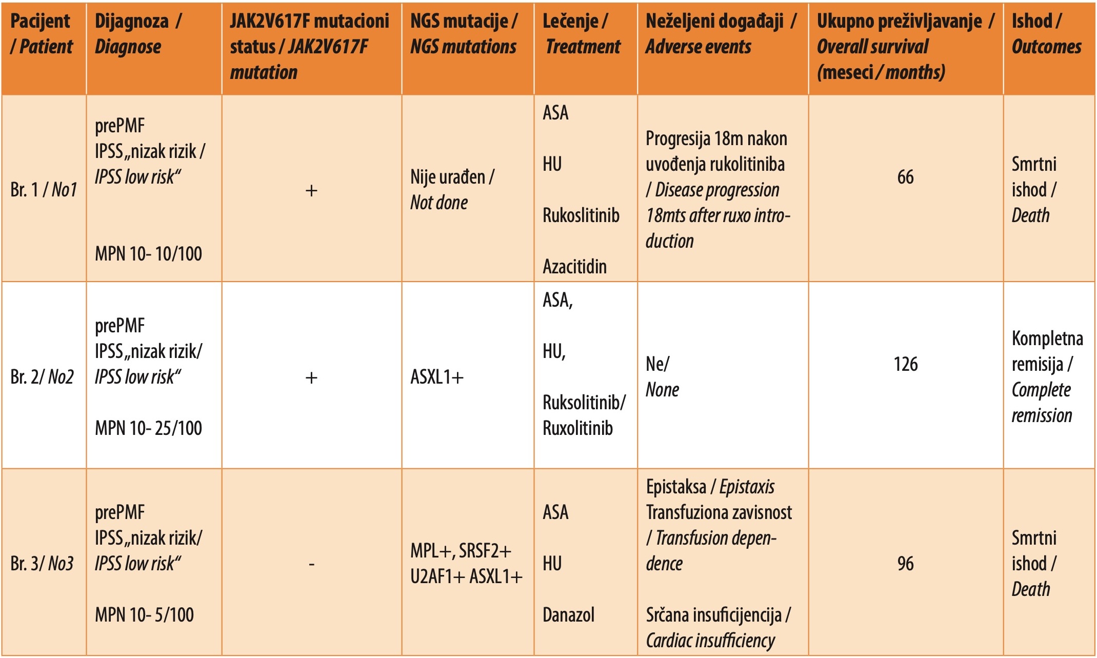
The clinical course of the disease in patients with lower-risk PMF shows unpredictability, which is confirmed by the previous and the most recent results of the “MOST” observational study, according to which 60% of patients (the median follow-up of a group of 232 patients with low/intermediate-1 risk MF was 52.9 months) have signs of disease progression, while 8.3% of patients had leukemic transformation or died [11],[17]. It should be noted that 84 patients had low-risk PMF, and the criteria for disease progression were Hb<100 g/L, Tr<100×109 /L, constitutional symptoms, the appearance or progression of splenomegaly, the presence of blasts >1% in peripheral blood, Le >25×109 /L, death due to disease progression, leukemic transformation and more than one transfusion Er. In our miniseries, a clear progression of the disease can be observed in all three cases, but with different therapeutic responses and clinical outcomes of the disease. Two patients had a fatal outcome: OS was 66 and 96 months, while the third was a patient in stable remission with an OS of 156 months. She is also a carrier of the ASXL1 mutation (Table 1).
CONCLUSION
Current data supports the significant presence of disease progression in patients with low-risk initial PMF, which requires regular monitoring of these patients with timely inclusion/change of current therapy in case of progression. Our data confirm that low-risk PMF is one entity with many different faces. Future research will show whether early NGS analysis of non-driver mutations and early therapy introduction contribute to changing the course of the disease and whether non-driver mutations will be a new target in targeted therapy for PMF.
Informations

-
Keywords:
-
Received:
-
Revised:
-
Accepted:
-
Online first:
-
DOI:
-
Cite this article:

Mihailo Smiljanić
Clinic for Hematology, University Clinical Center of Serbia
2 Dr Koste Todorovića Street, 11000 Belgrade, Serbia
E-mail:
1. England JT, McNamara CJ, Kennedy JA, Capo-Chichi JM, Huang J, Arruda A, et al. Clinical and molecular correlates of JAK-inhibitor therapy failure in myelofibrosis: long-term data from a molecularly annotated cohort. Leukemia. 2022 Jun;36(6):1689-92. doi: 10.1038/s41375-022-01544-x. [CROSSREF]
2. Li Y, Zhu S, Liu W, Ming J, Wang X, Hu X. Ruxolitinib-based combinations in the treatment of myelofibrosis: worth looking forward to. Ann Hematol. 2020 Jun;99(6):1161-76. doi: 10.1007/s00277-020-04028-z. [CROSSREF]
3. Tefferi A. Primary myelofibrosis: 2023 update on diagnosis, risk-stratification, and management. Am J Hematology. 2023 May;98(5):801-21. doi: 10.1002/ajh.26857. [CROSSREF]
4. Kvasnicka HM, Thiele J. Prodromal myeloproliferative neoplasms:the 2008 WHO classification. Am J Hematol. 2010 Jan;85(1):62-9. doi: 10.1002/ ajh.21543. [CROSSREF]
5. Tefferi A, Nicolosi M, Mudireddy M, Lasho TL, Gangat N, Begna KH, et al. Revised cytogenetic risk stratification in primary myelofibrosis: analysis based on 1002 informative patients. Leukemia. 2018 May;32(5):1189-99. doi: 10.1038/ s41375-018-0018-z. [CROSSREF]
6. Tefferi A, Pardanani A. Myeloproliferative neoplasms: a contemporary review. JAMA Oncol. 2015 Apr;1(1):97-105. doi: 10.1001/jamaoncol.2015.89. [CROSSREF]
7. Cervantes F, Dupriez B, Pereira A, Passamonti F, Reilly JT, Morra E, et al. New prognostic scoring system for primary myelofibrosis based on a study of the International Working Group for Myelofibrosis Research and Treatment. Blood. 2009 Mar 26;113(13):2895-901. doi: 10.1182/blood-2008-07-170449. [CROSSREF]
8. Passamonti F, Cervantes F, Vannucchi AM, Morra E, Rumi E, Pereira A, et al. A dynamic prognostic model to predict survival in primary myelofibrosis: a study by the IWG-MRT (International Working Group for Myeloproliferative Neoplasms Research and Treatment). Blood. 2010 Mar 4;115(9):1703-8. doi: 10.1182/blood-2009-09-245837. [CROSSREF]
9. Cervantes F. How I treat myelofibrosis. Blood 2014 Oct 23;124(17):2635-42. doi: 10.1182/blood-2014-07-575373. [CROSSREF]
10. Bose P. Management of patients with early myelofibrosis: a discussion of best practices. Curr Hematol Malig Rep. 2024 Jun;19(3):111-9. doi: 10.1007/ s11899-024-00729-8. [CROSSREF]
11. Gerds AT, Lyons RM, Colucci P, Kalafut P, Paranagama D, Verstovsek S. Disease and clinical characteristics of patients with a clinical diagnosis of myelofibrosis enrolled in the MOST study. Clin Lymphoma Myeloma Leuk. 2022 Jul;22(7):532-40. doi: 10.1016/j.clml.2022.02.001. [CROSSREF]
12. Al-Ali HK, Griesshammer M, Foltz L, Palumbo GA, Martino B, Palandri F, et al. Primary analysis of JUMP, a phase 3b, expanded-access study evaluating the safety and efficacy of ruxolitinib in patients with myelofibrosis, including those with low platelet counts. Br J Haematol. 2020 Jun;189(5):888-903. doi: 10.1111/bjh.16462. [CROSSREF]
13. Mead AJ, Milojkovic D, Knapper S, Garg M, Chacko J, Farquharson M, et al. Response to ruxolitinib in patients with intermediate-1-, intermediate-2-, and high-risk myelofibrosis: results of the UK ROBUST Trial. Br J Haematol. 2015 Jul;170(1):29-39. doi: 10.1111/bjh.13379. [CROSSREF]
14. Sørensen AL, Mikkelsen SU, Knudsen TA, Bjørn ME, Andersen CL, Bjerrum OW, et al. Ruxolitinib and interferon-α2 combination therapy for patients with polycythemia vera or myelofibrosis: a phase II study. Haematologica. 2020 Sep 1;105(9):2262-72. doi: 10.3324/haematol.2019.235648. [CROSSREF]
15. Shah S, Talati C, Al Ali N, Padron E, Sallman DA, Sweet KL, et al. Investigating patient characteristics and outcomes of myelofibrosis patients with hyperleukocytosis. Blood. 2018 Nov 29;132(Suppl 1):3041. doi: 10.1182/blood-2018-99-117834. [CROSSREF]
16. Yan X, Xu Z, Zhang P, Sun Q, Jia Y, Qin T, et al. Non-driver mutations landscape in different stages of primary myelofibrosis determined ASXL1 mutations play a critical role in disease progression. Blood Cancer J. 2023 Apr 20;13(1):56. doi: 10.1038/s41408-023-00829-3. [CROSSREF]
17. Komrokji RS, Grunwald MR, Braunstein E, Hamer-Maansson JE, Kalafut P, Mascarenhas J. Disease progression and leukemic transformation in patients with lower-risk myelofibrosis (MF): an analysis from MOST. Blood. 2022 Nov 15;140(Suppl 1):6832-4. doi: 10.1182/blood-2022-159181. [CROSSREF]