


Osteoporosis in autoimmune diseases
- University of Novi Sad, Faculty of Medicine, Novi Sad, Serbia
- University Clinical Center of Vojvodina, Clinic for Medical Rehabilitation, Novi Sad, Serbia
ABSTRACT
Introduction: Osteoporosis is one of the most common extra-articular complications in many chronic inflammatory rheumatic diseases, including rheumatoid arthritis, psoriatic arthritis, ankylosing spondylitis, and systemic lupus erythematosus etc..
Methods: We searched the PubMed, Scopus, and Web of Science databases for studies published within the last 10 years. This is a review article.
Results: Most studies on postmenopausal women have found a correlation between high levels of pro-inflammatory markers and increased bone loss. Literature data indicate a link between a low risk of major osteoporotic fractures, hip fractures, and non-vertebral fractures in patients with PsA who are on disease-modifying therapies. Patient assessment in autoimmune rheumatic inflammatory diseases includes medical history, clinical examination, fracture risk assessment, laboratory tests, and bone densitometry. The lack of adequate diagnosis and treatment of osteoporosis is influenced by factors such as insufficient knowledge about osteoporosis and its therapeutic benefits, concerns about potential side effects of treatment, low motivation, and inadequate patient education on the subject. Several methods have been developed to reduce prescribing errors and oversights, with the Fracture Liaison Service proving to be the most effective. Strategies for managing osteoporosis include patient education, lifestyle modifications, fall prevention, proper nutrition, disease activity control, and the introduction of osteoporosis medication.
Conclusion: Despite significant advances in prevention, diagnosis, and treatment, the prevalence of osteoporosis remains high and requires timely recognition and a more comprehensive approach. Assessing risk factors for falls and fractures, as well as recovery potential, is crucial in patients with autoimmune rheumatic inflammatory diseases.
INTRODUCTION
According to the World Health Organization (WHO) definition, osteoporosis is a systemic, progressive skeletal disease characterized by reduced bone mass and microarchitectural deterioration of bone tissue, resulting in increased bone fragility and susceptibility to fractures. Bone strength reflects the integration of two main characteristics: bone density and bone quality. Osteoporosis is a widespread condition, affecting one in three women and one in five men over the age of 50 years [1]. The population that is at greatest risk comprises postmenopausal women [2]. According to the results of a 2022 meta-analysis, the prevalence of osteoporosis and osteopenia ranges from 19.7% to 40.4% [3]. Among the most common risk factors for osteoporosis are advanced age, female sex, genetic predisposition, smoking, frequent alcohol consumption, inadequate nutrition, reduced intake of calcium and vitamin D, chronic use of systemic corticosteroids, as well as comorbidities, including autoimmune diseases, most commonly rheumatoid arthritis (RA) [4]. Osteoporosis represents a significant and often underrecognized complication in autoimmune diseases. The prevalence of autoimmune rheumatic diseases (ARDs) is estimated at 3,225 per 100,000 population [5]. Early recognition of these diseases is crucial for preventing complications and for initiating appropriate treatment [6].
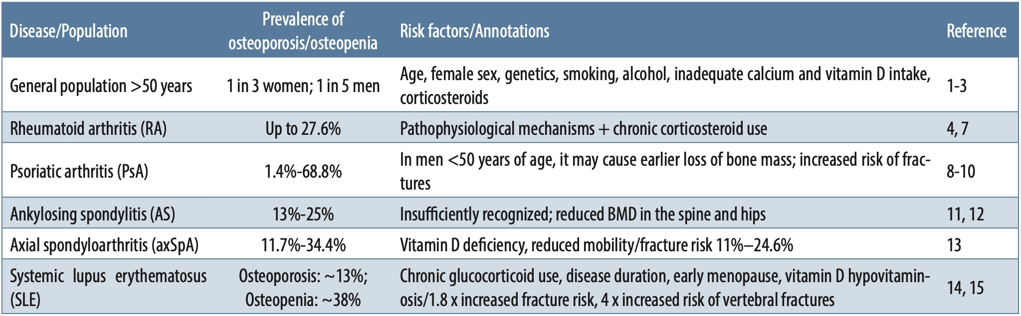
The prevalence of osteoporosis is significantly higher in patients with rheumatoid arthritis than in the general population, as much as by 27.6%, which can be explained by the pathophysiological mechanisms of the disease as well as by therapeutic approaches in rheumatoid arthritis, which very often include the chronic use of systemic corticosteroid therapy [7]. The results of an analysis of 21 published studies indicate that the prevalence of osteoporosis in patients with psoriatic arthritis (PsA) ranges from 1.4% to 68.8% [8]. According to data from a retrospective study published in 2024, the incidence of osteoporosis and osteopenia in patients with psoriatic arthritis ranges between 11.7% and 33.1% [9]. In males younger than 50 years, PsA may contribute to earlier bone mass loss compared with traditional risk factors [9]. Patients with psoriatic arthritis also have an increased fracture risk, as compared with the general population [10].
Osteoporosis is a common condition in patients with ankylosing spondylitis (AS) but remains underrecognized in clinical practice [11]. A combined assessment using dual-energy X-ray absorptiometry (DXA) and quantitative computed tomography revealed reduced bone mineral density (BMD) and bone loss in the spine and hips of patients with early and long-standing AS [12]. The prevalence of osteoporosis in patients with axial spondyloarthritis (axSpA) ranges from 11.7% to 34.4%, while the fracture risk ranges from 11% to 24.6%, with significant risk factors including vitamin D deficiency and reduced patient mobility due to the underlying disease [13].
The prevalence of osteoporosis in patients with systemic lupus erythematosus (SLE) is approximately 13%, while the prevalence of osteopenia is approximately 38% [14]. The main risk factors in this population include chronic glucocorticoid use, disease duration, and early menopause associated with reduced bone mineral density (BMD) and vitamin D hypovitaminosis [15]. Patients with SLE are at a 1.8 times higher overall fracture risk and at a four times higher risk of vertebral fractures [14]. The results of the above-described studies are presented in Table 1. The table summarizes the prevalence of osteoporosis and osteopenia in the general population and among patients with various inflammatory rheumatic diseases, highlighting key risk factors and specific annotations for each group.
MATERIALS AND METHODS
The methodology of this study is based on a literature review of the PubMed, Scopus, and Web of Science databases, using keywords related to rheumatic diseases and osteoporosis (osteoporosis, prevalence, risk factors, fractures, therapy). Articles published in English within the last ten years focusing on adult patients and reporting data on prevalence, risk factors, and therapeutic approaches were included. Studies that were not relevant, those involving pediatric populations, and case reports were excluded. The collected data were analyzed descriptively and thematically, with a focus on clinical characteristics, mechanisms of bone loss, and treatment strategies. This is a review article.
Factors affecting bone mineral density in autoimmune rheumatic diseases
Chronic inflammatory responses in autoimmune rheumatic diseases (ARDs), such as rheumatoid arthritis and systemic lupus erythematosus, activate osteoclasts, thereby accelerating bone resorption and leading to reduced bone density [16]. Cytokines released by immune cells during the inflammatory response affect bone metabolism, promoting the development of osteoporosis. The formation of syndesmophytes and ankylosis further increases mechanical load in cortical bone regions and paravertebral ligaments, while the loss of trabecular bone in vertebral bodies can result in decreased bone mineral density [17]. Pain and reduced mobility caused by these diseases limit patients’ physical activity, which further adversely affects bone health [18].
A possible link between psoriatic arthritis (PsA) and osteoporosis may be found in the more pronounced demineralization observed in patients with PsA, which is intensified by aging, estrogen deficiency, and impaired quality-of-life indexes [19]. Studies have shown that patients with PsA are more likely to develop fractures compared with a healthy control group without psoriasis; however, a definite association between a high fracture risk and reduced bone mineral density has not been conclusively established. Further research is needed to confirm PsA as a risk factor for osteoporosis [20]. More recent literature data have shown that in patients with PsA, men have an increased risk of osteoporosis as well as of osteoporotic fractures, even when they are younger than 50 years [21]. PsA has been described as an independent risk factor for reduced bone density and falls, and consequently for associated fractures [22].
Patients with ankylosing spondylitis (AS) have reduced bone mineral density (BMD) and an increased fracture risk. Younger patients and those in the early stages of the disease are at additional risk of osteoporosis and fractures [23]. Vertebral fractures in AS are often unstable due to ossification of supporting and elastic ligaments. Inflammatory cytokines, including tumor necrosis factor (TNF)-alpha and interleukins (IL) such as IL-1, IL-6, and IL-17, increase osteoclast activity and bone resorption [24]. Studies indicate that bone loss and fractures are associated with systemic lupus erythematosus (SLE), which is associated not only with the disease itself but also with low vitamin D levels and adverse effects of therapy [25].
The effect of medications used in the treatment of autoimmune rheumatic diseases on bone mineral density
Medications used in the treatment of autoimmune rheumatic diseases are essential for managing the underlying condition but have numerous adverse effects on the musculoskeletal system and bone density. Owing to its anti-inflammatory effects, glucocorticoid therapy is frequently used in patients with autoimmune rheumatic diseases (ARDs). Understanding the mechanisms of action of glucocorticoids on bone, the immunological interactions, and the role of vitamin D, as well as recognizing the impact of chronic inflammation on bone metabolism, enables a better understanding of their adverse skeletal effects [26]. Glucocorticoids reduce calcium absorption in the gastrointestinal tract, increase renal calcium excretion, and suppress osteoblast function, all of which lead to decreased bone tissue synthesis. They also promote apoptosis of osteocytes and osteoblasts, resulting in increased bone resorption. Together, these effects contribute to a reduction in BMD and an increased fracture risk [27]. The fracture risk, particularly for vertebral fractures, increases significantly after continuous glucocorticoid use for three months and continues to rise with longer duration of therapy [28]. Daily corticosteroid doses exceeding 7.5 mg increase the risk of major osteoporotic fractures by 15% and the risk of hip fractures by 20% [29]. To mitigate these adverse effects, it is recommended to use the lowest effective therapeutic doses of glucocorticoids for the shortest possible duration. Concomitant administration of vitamin D and calcium is necessary to reduce the harmful effects of glucocorticoids on bone tissue [29].
Conventional disease-modifying drugs (disease-modifying antirheumatic drugs, DMARDs), including methotrexate and sulfasalazine, which are routinely used in patients with ARDs, have a lesser impact on BMD compared with glucocorticoids. In some studies, methotrexate has been associated with reduced bone matrix synthesis; however, the reasons for this association have not yet been fully clarified. Patients receiving methotrexate therapy may have an increased fracture risk, which is attributed not to the medication itself but to the underlying disease and other risk factors [30].
Biological therapy (TNF inhibitors and IL inhibitors) targets specific components of the immune response to suppress inflammatory processes in the body. By reducing systemic inflammation, these agents have a positive effect on BMD and decrease bone resorption in patients with RA and other ARDs [31]. Janus kinase (JAK) inhibitors, such as tofacitinib and baricitinib, represent a newer group of disease-modifying agents used in the treatment of ARDs. Their impact on BMD is not yet fully known. Based on currently available data, they are considered to have a neutral or even potentially positive effect on bone metabolism [32]. Hormone replacement therapy (HRT) plays an important role in autoimmune diseases. In hypothyroidism induced by autoimmune thyroiditis, thyroid hormone replacement maintains a euthyroid state, reduces hypothyroid symptoms, and contributes to the preservation of bone mass. This reduces the risk of osteoporosis and fractures, which are common problems in patients with chronic autoimmune disorders [33].
DIAGNOSTICS
The gold standard for diagnosing osteoporosis is dual-energy X-ray absorptiometry (DXA), which measures bone mineral density (BMD). The diagnostic process begins with a clinical assessment of risk factors, including age, sex, previous fractures, family history of osteoporosis, glucocorticoid use, and lifestyle factors [34],[35]. After identifying patients at risk, DXA examination of the lumbar spine and femoral neck is performed. The main criterion required for establishing a diagnosis of osteoporosis is a T-score < −2.5 [36]. However, diagnostic criteria are not based solely on the T-score. The fracture risk must also be taken into account, as assessed on the basis of previous fractures, comorbidities, and other risk factors [37]. The Z-score is used in younger individuals and perimenopausal women; values ≤ −2 indicate BMD lower than expected for the patient’s age [38]. Clinical guidelines recommend performing DXA in all women older than 65 years, as well as in younger postmenopausal women and men older than 50 years with additional risk factors [38].
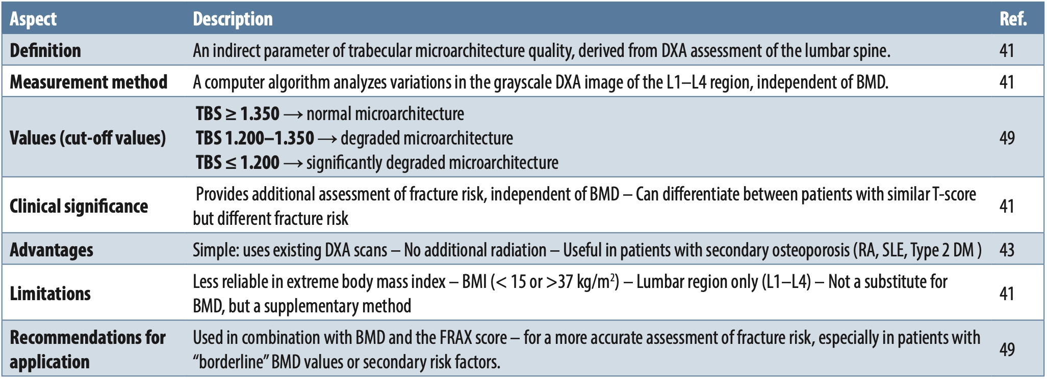
Additional laboratory diagnostics include assessment of bone metabolism parameters (parathyroid hormone, N-terminal propeptide of type I procollagen, bone alkaline phosphatase, beta-CrossLaps, phosphorus, vitamin D, and calcium), as well as evaluation of thyroid and parathyroid gland function. The Fracture Risk Assessment Tool (FRAX) is a useful online tool that calculates the 10-year probability of hip fracture or other major osteoporotic fractures [39]. An enhanced version, FRAX Plus, incorporates additional factors such as the location of previous fractures, the number of falls in the preceding year, corticosteroid doses, and the trabecular bone score (TBS), thereby improving the accuracy of risk assessment [40]. Interpretation of TBS values is presented in Table 2. The table summarizes key aspects of TBS: what it measures (bone microarchitecture), how it is measured (analysis of lumbar spine DXA images), clinical significance (complement to BMD in fracture risk assessment), advantages, limitations, and recommended use.
Certain diagnostic criteria routinely used in the general population may be unreliable in patients with autoimmune rheumatic diseases. In patients with ARDs (RA, axSpA, PsA, SLE), it is necessary to adapt the diagnostic approach because both the disease itself and its treatment may affect DXA measurement results. Localized bone loss in RA and new bone formation in axSpA may lead to falsely low or falsely elevated BMD values, making interpretation more challenging [41].
In such cases, additional assessment of peripheral bone density, such as BMD of the distal forearm, as well as the use of TBS, may be useful for a more accurate evaluation of fracture risk. In patients with axSpA and a high fracture risk, combining DXA and TBS methods is recommended to better identify those who require early intervention [42]. Timely assessment and continuous monitoring are of crucial importance, particularly in patients receiving long-term glucocorticoid therapy, in whom more frequent BMD monitoring and reassessment of fracture risk are recommended [43].
The application of the FRAX algorithm in patients with autoimmune rheumatic diseases has particular clinical significance, as it enables early identification of individuals at increased fracture risk and facilitates decisions regarding the initiation of specific therapy. Inclusion of data on glucocorticoid use in the FRAX index significantly contributes to a more accurate fracture risk assessment, given that chronic glucocorticoid therapy is one of the leading causes of secondary osteoporosis [41]. When the measured BMD value at the femoral neck is also entered into FRAX, risk estimation becomes even more reliable, which is particularly important in patients with RA or axSpA, in whom osteoporosis may occur at a younger age [44].
However, it is important to emphasize that FRAX does not take into account disease activity, the degree of inflammation, functional disability, or the frequency of falls, and therefore may underestimate risk in patients with highly active disease or those with significant structural damage. In addition, the standard FRAX model does not differentiate between low and high doses of glucocorticoids. Thus, in patients receiving long-term therapy with doses exceeding 7.5 mg of prednisone, adjustment of the calculated risk by an additional 15-20% is recommended [45].
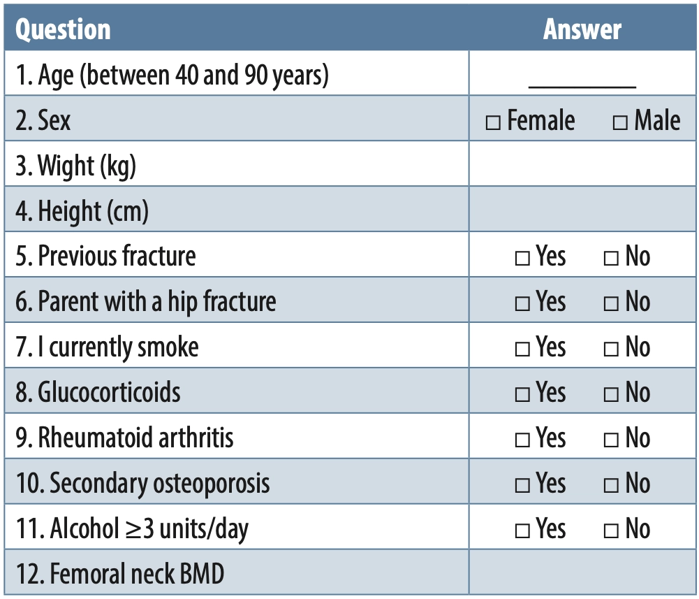
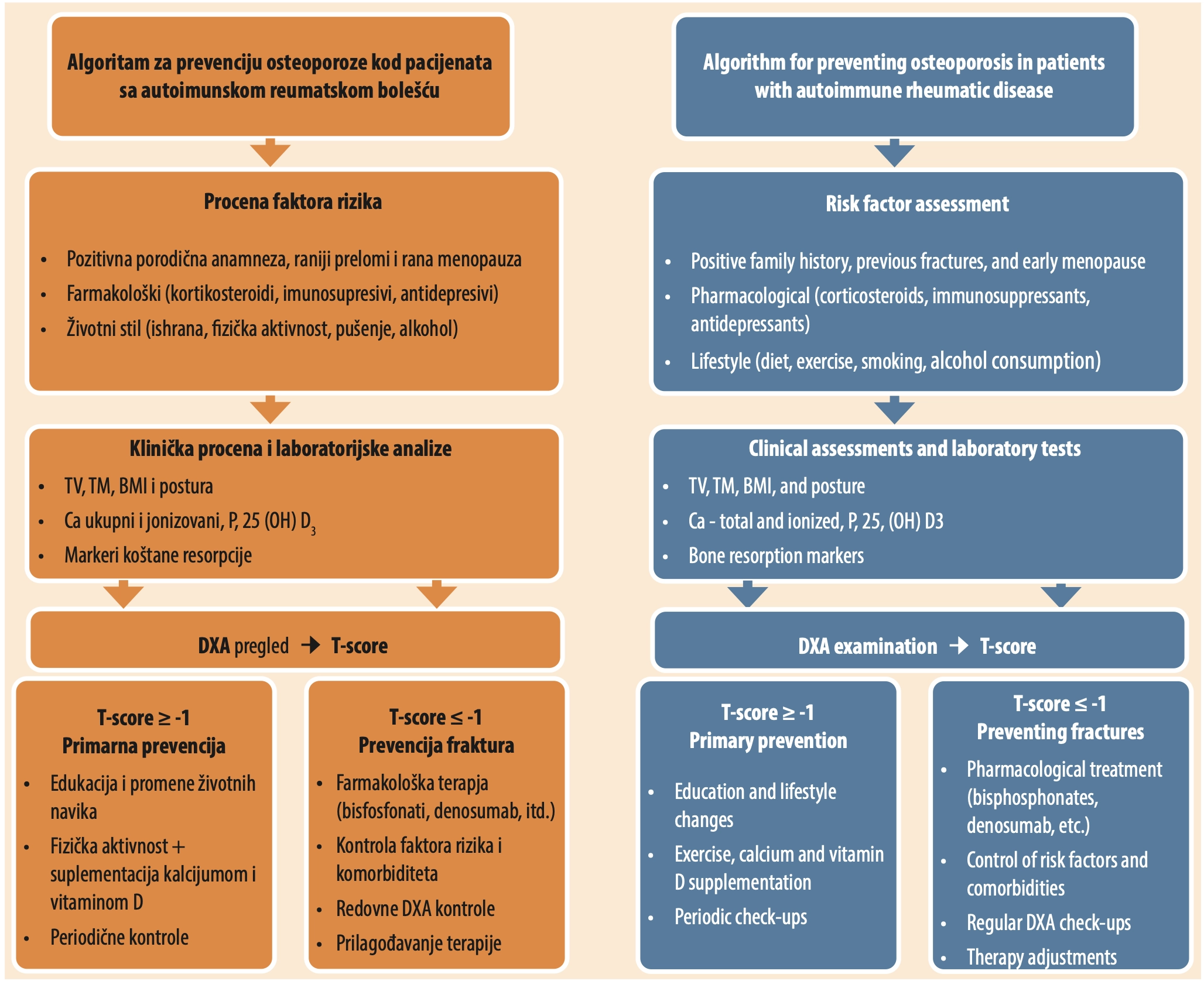
Because of these limitations, combining the FRAX tool with additional assessment methods, such as TBS and vertebral morphometry, is recommended in order to improve identification of patients at high fracture risk and optimize therapeutic decision-making. Vertebral morphometry (lateral vertebral assessment – LVA) is an imaging method that uses DXA to detect and analyze vertebral fractures. Unlike conventional radiography, LVA allows for lower radiation exposure and faster assessment of vertebral deformities. In the context of osteoporosis and autoimmune diseases, LVA is particularly useful because detecting fractures enables timely adjustment of therapy and a more accurate assessment of the risk of subsequent fractures [46]. Table 3 and Figure 1 presents the FRAX questionnaire. Figure 1 presents the algorithm designed to assist in the prevention and treatment of osteoporosis, according to the literature.
 33],[34],[36]" width="2056" height="1676">
CLINICAL PRACTICE GUIDELINES
Timely identification of risk factors, patient education, and initiation of appropriate therapy can significantly reduce fracture risk and improve quality of life. Prevention includes modification of risk factors, calcium and vitamin D supplementation, appropriately tailored physical activity, regular monitoring of bone density, and fall prevention strategies [33].
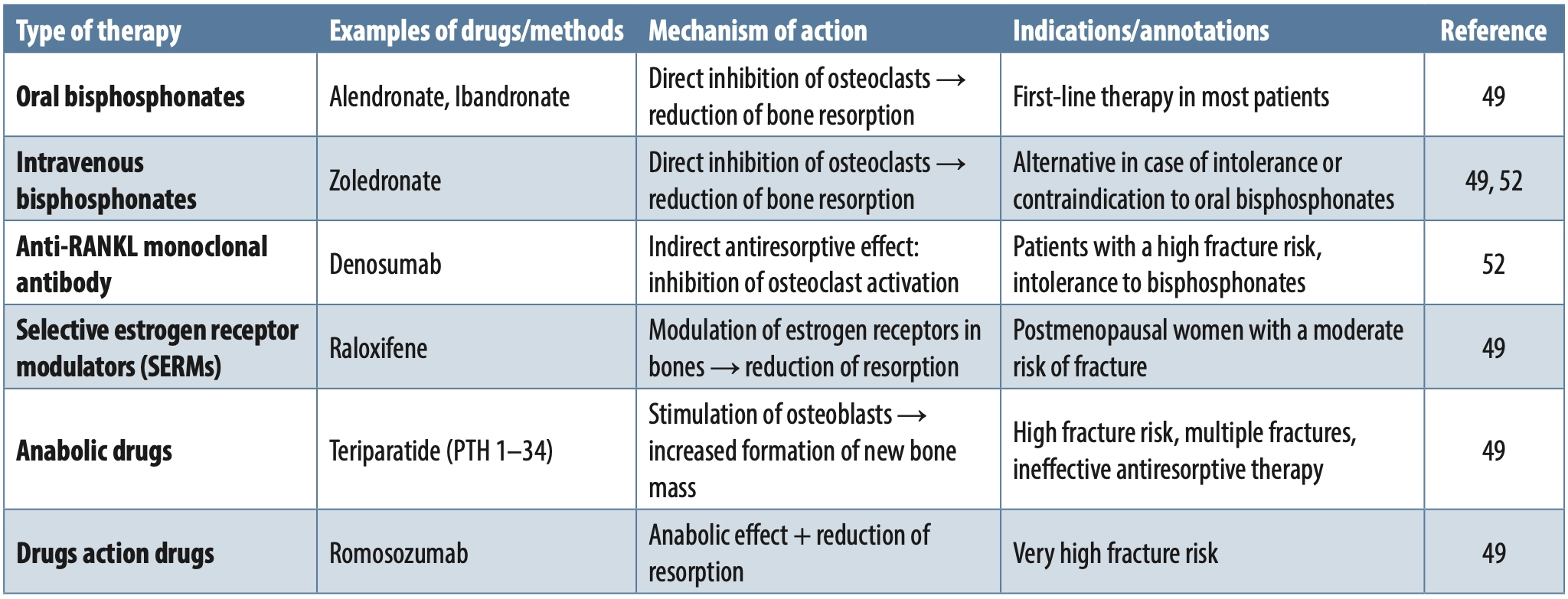
First-line therapy for the treatment of osteoporosis in most patients is based on the use of antiresorptive agents, primarily oral bisphosphonates, in accordance with the recommendations of the American Association of Clinical Endocrinology (AACE) [47]. In patients who are intolerant of or have contraindications to oral bisphosphonates, intravenous bisphosphonates or denosumab are alternative options. Both categories of medications belong to the group of antiresorptive drugs. Bisphosphonates, regardless of the route of administration (oral or intravenous), directly inhibit osteoclast activity, whereas denosumab, a monoclonal antibody that binds to the receptor activator of nuclear factor kappa-B ligand (RANKL), exerts its antiresorptive effect indirectly [48]. Selective estrogen receptor modulators (SERMs) may be considered as an additional option, particularly in postmenopausal women who are at moderate risk. For patients at high risk of fracture who are unable to use oral therapy, treatment with denosumab or teriparatide (a recombinant form of parathyroid hormone), administered subcutaneously, is recommended. In patients at very high risk of fracture or those who have already sustained multiple fractures, initiation of anabolic agents (teriparatide) or agents with a dual mechanism of action, such as romosozumab, is recommended. Romosozumab is a monoclonal antibody that inhibits sclerostin, thereby promoting new bone formation and reducing osteoclast activity. It is administered subcutaneously once a year. It is important to emphasize that fracture risk should be reassessed after each new fracture, regardless of the time of occurrence, and that fracture risk increases after discontinuation of therapy [49].
In patients with rheumatoid arthritis (RA), adequate treatment of the underlying disease plays an additional role in bone protection. Conventional disease-modifying antirheumatic drugs (e.g., methotrexate) and biological agents (TNF inhibitors, IL-6 inhibitors, rituximab, abatacept) reduce disease activity and thereby decrease inflammation, which may have a beneficial effect on bone metabolism, although their direct impact on BMD is limited [50].
In adults who are beginning or continuing glucocorticoid therapy for longer than three months, the recommendations of the American College of Rheumatology (ACR) emphasize the need for an initial fracture risk assessment immediately after beginning treatment. In individuals older than 40 years, this assessment includes clinical evaluation of prior fractures, DXA measurement with lateral vertebral assessment (LVA) or spinal radiography, as well as calculation of the FRAX risk. In patients with moderate, high, or very high fracture risk, initiation of pharmacological therapy – oral or intravenous bisphosphonates, denosumab, or teriparatide – is strongly recommended. In patients with hormone deficiencies, substitution therapy may also contribute to the modulation of the immune response and the reduction of the risk of complications associated with autoimmune diseases [50]. These recommendations are presented in Table 4 with the corresponding references. This table presents different treatments for osteoporosis, including examples of drugs, their mechanisms of action, and indications. It covers oral and intravenous bisphosphonates, RANKL monoclonal antibodies, selective estrogen receptor modulators (SERMs), and anabolic drugs such as teriparatide and romosozumab, along with guidelines on when each treatment is used.
Fracture Liaison Services (FLS) represent an organized care model whose primary goal is to reduce fracture risk in individuals older than 50 years through timely diagnosis and initiation of appropriate osteoporosis treatment. Recent studies show that implementation of FLS protocols has led to a significant reduction in fracture risk in both sexes (by 13% in women and by 10% in men), as well as a reduction in mortality rates by 18% in women and by 15% in men [51]. The introduction of the FLS approach in patients with autoimmune rheumatic diseases is of particular importance, as it enables timely diagnosis, early initiation of therapy, and a multidisciplinary approach involving a rheumatologist, endocrinologist, and physiatrist [52].
CONCLUSION
Osteoporosis is a common and serious complication of autoimmune rheumatic diseases that significantly affects patients’ quality of life and functional status. Its development is multifactorial: chronic inflammation and immune dysregulation promote osteoclast activation and accelerated bone resorption, while pharmacological treatments, particularly long-term glucocorticoid therapy, further inhibit osteoblast function and reduce bone mineral density. In addition, limited mobility and reduced physical activity in patients with pain and deformities contribute to the progression of osteoporosis.
Prevention of osteoporosis represents the cornerstone of preserving bone mass and reducing fracture risk in patients with autoimmune rheumatic diseases. Key preventive approaches include appropriately tailored physical activity, ensuring adequate intake of calcium and vitamin D, and reducing risk factors related to pharmacological therapy and lifestyle. Timely identification of patients at increased risk through clinical assessment of risk factors, DXA scanning, the FRAX index, and additional methods, such as TBS, enables early implementation of preventive measures and personalized follow-up. When prevention is insufficient, and osteoporosis progresses, targeted pharmacological treatment – taking into account the type and activity of the underlying disease – can significantly reduce the fracture risk and preserve patients’ functional capacity. Pharmacological strategies, including antiresorptive and anabolic agents, should be individualized, taking into account the type and activity of the underlying disease, patient age, and the presence of comorbidities. Integration of therapy for the underlying disease with specific osteoporosis treatment significantly reduces fracture risk and contributes to the preservation of functional capacity.
A multidisciplinary approach is essential for optimizing strategies of prevention, monitoring, and treatment. Future research should focus on targeted therapies that reduce bone loss without compromising immune control, as well as on improving risk assessment tools to better identify patients at high risk of developing osteoporosis. By treating osteoporosis as an integral component of the management of autoimmune diseases, clinicians can reduce the fracture risk and improve the long-term quality of life for affected patients.
Informations

-
Keywords:
-
Received:
-
Revised:
-
Accepted:
-
Online first:
-
DOI:
-
Cite this article:

Sandra Jelčić
Clinic for Medical Rehabilitation, University Clinical Center of Vojvodina
1 Hajduk Veljkova Street, 21000 Novi Sad, Serbia
E-mail:
1. Kao FC, Hsu YC, Tu YK, Chen TS, Wang HH, Lin JC. Long-Term Use of Immunosuppressive Agents Increased the Risk of Fractures in Patients with Autoimmune Diseases: An 18-Year Population-Based Cohort Study. Biomed. 2023 Oct 12;11(10):2764. doi: 10.3390/biomedicines11102764. [CROSSREF]
2. Aibar-Almazán A, Voltes-Martínez A, Castellote-Caballero Y, Afanador-Restrepo DF, Carcelén-Fraile MDC, López-Ruiz E. Current Status of the Diagnosis and Management of Osteoporosis. Int J Mol Sci. 2022 Aug 21;23(16):9465. doi: 10.3390/ijms23169465. [CROSSREF]
3. Xiao PL, Cui AY, Hsu CJ, Peng R, Jiang N, Xu XH, et al. Global, regional prevalence, and risk factors of osteoporosis according to the World Health Organization diagnostic criteria: a systematic review and meta-analysis. Osteoporos Int. 2022 Oct;33(10):2137-53. doi: 10.1007/s00198-022-06454-3. [CROSSREF]
4. Compston JE, McClung MR, Leslie WD. Osteoporosis. Lancet. 2019 Jan 26;393(10169):364-76. doi: 10.1016/S0140-6736(18)32112-3. [CROSSREF]
5. Miller FW. The increasing prevalence of autoimmunity and autoimmune diseases: an urgent call to action for improved understanding, diagnosis, treatment, and prevention. Curr Opin Immunol. 2023 Feb;80:102266. doi: 10.1016/j.coi.2022.102266. [CROSSREF]
6. Lenti MV, Rossi CM, Melazzini F, Gastaldi M, Bugatti S, Rotondi M, et al. A Seronegative autoimmune disease: A challenging diagnosis. Autoimmun Rev. 2022 Sep;21(9):103143. doi: 10.1016/j.autrev.2022.103143. [CROSSREF]
7. Moshayedi S, Tasorian B, Almasi-Hashiani A. The prevalence of osteoporosis in rheumatoid arthritis patient: a systematic review and meta-analysis. Sci Rep. 2022 Sep 23;12(1):15844. doi: 10.1038/s41598-022-20016-x. [CROSSREF]
8. Chandran S, Aldei A, Johnson SR, Cheung AM, Salonen D, Gladman DD. Prevalence and risk factors of low bone mineral density in psoriatic arthritis: A systematic review. Semin Arthritis Rheum. 2016 Oct;46(2):174-82. doi: 10.1016/j.semarthrit.2016.05.005. [CROSSREF]
9. Takami K, Higashiyama M, Tsuji S. Osteoporosis and osteopenia in patients with psoriatic arthritis: A single-centre retrospective study. Mod Rheumatol. 2024 Oct 15;34(6):1252-7. doi: 10.1093/mr/roae028. [CROSSREF]
10. Martínez-Vidal MP, Jovani V, Noguera-Pons JR, Álvarez-Cienfuegos A. Osteoporosis in psoriatic arthritis: Risk factors, insufficiency fractures and its association with the disease activity. Reumatol Clin (Engl Ed). 2024 Jan;20(1):8- 13. doi: 10.1016/j.reumae.2023.05.007. [CROSSREF]
11. Sharif K, Tsur AM, Ben-Shabat N, Watad A, Cohen AD, Amital H. The risk of osteoporosis in patients with ankylosing spondylitis-A large retrospective matched cohort study. Med Clin (Barc). 2023 May 12;160(9):373-8. doi: 10.1016/j.medcli.2022.10.013. [CROSSREF]
12. Zheng B, Yu P, Liu H, Liang Y. Anti-Osteoporosis Is Imperative in Prevention of Progress of Ankylosing Spondylitis. Int J Gen Med. 2025 Jan 21;18:291-7. doi: 10.2147/IJGM.S509296. [CROSSREF]
13. Ramírez J, Nieto-González JC, Curbelo Rodríguez R, Castañeda S, Carmona L. Prevalence and risk factors for osteoporosis and fractures in axial spondyloarthritis: A systematic review and meta-analysis. Semin Arthritis Rheum. 2018 Aug;48(1):44-52. doi: 10.1016/j.semarthrit.2017.12.001. [CROSSREF]
14. Lai SW, Kuo YH, Liao KF. Bone health in patients with systemic lupus erythematosus. Ann Rheum Dis. 2021 Nov;80(11):e179. doi: 10.1136/annrheumdis-2019-216417. [CROSSREF]
15. Gu C, Zhao R, Zhang X, Gu Z, Zhou W, Wang Y, et al. A meta-analysis of secondary osteoporosis in systemic lupus erythematosus: prevalence and risk factors. Arch Osteoporos. 2019 Dec 4;15(1):1. doi:10.1007/s11657-019-0667-1. [CROSSREF]
16. Hong CX, Pan YZ, Dai FB. Potential association of rheumatic diseases with bone mineral density and fractures: a bi-directional mendelian randomization study. BMC Musculoskelet Disord. 2024 Jul 5;25(1):521. doi: 10.1186/s12891-024-07496-w. [CROSSREF]
17. Kim JW, Park S, Jung JY, Kim HA, Kwon SR, Choi ST, et al. Prevalence and Factors of Osteoporosis and High Risk of Osteoporotic Fracture in Patients with Ankylosing Spondylitis: A Multicenter Comparative Study of Bone Mineral Density and the Fracture Risk Assessment Tool. J Clin Med. 2022 May 17;11(10):2830. doi: 10.3390/jcm11102830. [CROSSREF]
18. Kwok TSH, Sutton M, Ye JY, Pereira D, Chandran V, Gladman DD. Prevalence and Factors Associated with Osteoporosis and Bone Mineral Density Testing in Psoriatic Arthritis. Arthritis Care Res (Hoboken). 2022 Jun;74(6):1006-12. doi: 10.1002/acr.24538. [CROSSREF]
19. Chen TL, Lu JW, Huang YW, Wang JH, Su KY. Bone Mineral Density, Osteoporosis, and Fracture Risk in Adult Patients with Psoriasis or Psoriatic Arthritis: A Systematic Review and Meta-Analysis of Observational Studies. J Clin Med. 2020 Nov 19;9(11):3712. doi: 10.3390/jcm9113712. [CROSSREF]
20. Schauer A, Uthayakumar AK, Boardman G, Bunker CB. The risk of osteopenia/osteoporosis and psoriatic disease: A systematic review. Skin Health Dis. 2022 Sep 21;3(1):e169. doi: 10.1002/ski2.169. [CROSSREF]
21. Halasi A, Szegedi A, Törőcsik D, Varga J, Farmasi N, Szűcs G, et al. Psoriatic arthritis and its special features predispose not only for osteoporosis but also for fractures and falls. J Dermatol. 2023 May;50(5):608-14. doi: 10.1111/1346-8138.16710. [CROSSREF]
22. Mei J, Hu H, Ding H, Huang Y, Zhang W, Chen X, et al. Investigating the causal relationship between ankylosing spondylitis and osteoporosis in the European population: a bidirectional Mendelian randomization study. Front Immunol. 2023 Jun 8;14:1163258. doi: 10.3389/fimmu.2023.1163258. [CROSSREF]
23. Yan F, Wu L, Lang J, Huang Z. Bone density and fracture risk factors in ankylosing spondylitis: a meta-analysis. Osteoporos Int. 2024 Jan;35(1):25-40. doi: 10.1007/s00198-023-06925-1. [CROSSREF]
24. Rella V, Rotondo C, Altomare A, Cantatore FP, Corrado A. Bone Involvement in Systemic Lupus Erythematosus. Int J Mol Sci. 2022 May 22;23(10):5804. doi: 10.3390/ijms23105804. [CROSSREF]
25. Salman-Monte TC, Torrente-Segarra V, Vega-Vidal AL, Corzo P, Castro-Dominguez F, Ojeda F, et al. Bone mineral density and vitamin D status in systemic lupus erythematosus (SLE): A systematic review. Autoimmun Rev. 2017 Nov;16(11):1155-9. doi: 10.1016/j.autrev.2017.09.011. [CROSSREF]
26. Cho SK, Sung YK. Update on Glucocorticoid Induced Osteoporosis. Endocrinol Metab (Seoul). 2021 Jun;36(3):536-43. doi: 10.3803/EnM.2021.1021. [CROSSREF]
27. Zerikly R, Demetriou EW. Use of Fracture Risk Assessment Tool in clinical practice and Fracture Risk Assessment Tool future directions. Womens Health (Lond). 2024 Jan-Dec;20:17455057241231387. doi: 10.1177/17455057241231387. [CROSSREF]
28. Hayes KN, Baschant U, Hauser B, Burden AM, Winter EM. When to Start and Stop Bone-Protecting Medication for Preventing Glucocorticoid-Induced Osteoporosis. Front Endocrinol (Lausanne). 2021 Dec 15;12:782118. doi: 10.3389/fendo.2021.782118. Erratum in: Front Endocrinol (Lausanne). 2022 Mar 14;13:877512. doi: 10.3389/fendo.2022.877512. [CROSSREF]
29. Zerbini CAF, Clark P, Mendez-Sanchez L, Pereira RMR, Messina OD, Uña CR, et al. Chronic Inflammation and Bone Structure (CIBS) Working Group. Biologic therapies and bone loss in rheumatoid arthritis. Osteoporos Int. 2017 Feb;28(2):429-46. doi: 10.1007/s00198-016-3769-2. [CROSSREF]
30. Lv F, Hu S, Lin C, Cai X, Zhu X, Ji L. Association between biologic therapy and fracture incidence in patients with selected rheumatic and autoimmune diseases: A systematic review and meta-analysis of randomized controlled trials. Pharmacol Res. 2022 Jul;181:106278. doi: 10.1016/j.phrs.2022.106278. [CROSSREF]
31. Hu L, Liu R, Zhang L. Advance in bone destruction participated by JAK/STAT in rheumatoid arthritis and therapeutic effect of JAK/STAT inhibitors. Int Immunopharmacol. 2022 Oct;111:109095. doi: 10.1016/j.intimp.2022.109095. [CROSSREF]
32. Wu J, Huang H, Yu X. How does Hashimoto’s thyroiditis affect bone metabolism? Rev Endocr Metab Disord. 2023 Apr;24(2):191-205. doi: 10.1007/s11154-022-09778-x. [CROSSREF]
33. Bjelica A, Vučaj Ćirilović V, Tomašević Todorović S, Filipović K. Postmenopausal osteoporosis. Med Pregl. 2018 May-Jun;71(5-6):201-5. doi: 10.2298/ MPNS1806201B. [CROSSREF]
34. Tomasevic-Todorovic S, Vazic A, Issaka A, Hanna F. Comparative assessment of fracture risk among osteoporosis and osteopenia patients: a cross-sectional study. Open Access Rheumatol. 2018 May 30;10:61-6. doi: 10.2147/OARRR.S151307. [CROSSREF]
35. Morin SN, Feldman S, Funnell L, Giangregorio L, Kim S, McDonald-Blumer H, et al.; Osteoporosis Canada 2023 Guideline Update Group. Clinical practice guideline for management of osteoporosis and fracture prevention in Canada: 2023 update. CMAJ. 2023 Oct 10;195(39):E1333-48. doi: 10.1503/cmaj.221647. [CROSSREF]
36. Tomasević-Todorović S, Simić-Panić D, Knežević A, Demeši-Drljan Č, Marić D, Hanna F. Osteoporosis in patients with stroke: A cross-sectional study. Ann Indian Acad Neurol. 2016 Apr-Jun;19(2):286-8. doi: 10.4103/0972-2327.173409. [CROSSREF]
37. Arceo-Mendoza RM, Camacho PM. Postmenopausal Osteoporosis: Latest Guidelines. Endocrinol Metab Clin North Am. 2021 Jun;50(2):167-78. doi: 10.1016/j.ecl.2021.03.009. [CROSSREF]
38. Kanis JA, Johansson H, Harvey NC, Gudnason V, Sigurdsson G, Siggeirsdottir K, et al. Adjusting conventional FRAX estimates of fracture probability according to the recency of sentinel fractures. Osteoporos Int. 2020 Oct;31(10):1817-28. doi: 10.1007/s00198-020-05517-7. [CROSSREF]
39. Kanis JA, Johansson H, Harvey NC, Lorentzon M, Liu E, Vandenput L, et al. Adjusting conventional FRAX estimates of fracture probability according to the number of prior falls in the preceding year. Osteoporos Int. 2023 Mar;34(3):479-87. doi: 10.1007/s00198-022-06633-2. [CROSSREF]
40. Johansson H, Kanis JA, Odén A, Leslie WD, Fujiwara S, Glüer CC, et al. Impact of femoral neck and lumbar spine BMD discordances on FRAX probabilities in women: a meta-analysis of international cohorts. Calcif Tissue Int. 2014 Nov;95(5):428-35. doi: 10.1007/s00223-014-9911-2. [CROSSREF]
41. McCloskey EV, Odén A, Harvey NC, Leslie WD, Hans D, Johansson H, et al. A Meta-Analysis of Trabecular Bone Score in Fracture Risk Prediction and Its Relationship to FRAX. J Bone Miner Res. 2016 May;31(5):940-8. doi: 10.1002/ jbmr.2734. [CROSSREF]
42. Puksun K, Pongchaiyakul C, Pakchotanon R, Narongroeknawin P, Leosuthamas P, Arunthanachaikul T, et al. Comparison of different intervention thresholds for the treatment of glucocorticoid-induced osteoporosis: a cross-sectional study. BMC Rheumatol. 2025 Apr 2;9(1):38. doi: 10.1186/ s41927-025-00488-w. [CROSSREF]
43. Rotta D, Fassio A, Rossini M, Giollo A, Viapiana O, Orsolini G, et al. Osteoporosis in Inflammatory Arthritides: New Perspective on Pathogenesis and Treatment. Front Med (Lausanne). 2020 Dec 1;7:613720. doi: 10.3389/ fmed.2020.613720. [CROSSREF]
44. Kanis JA, Johansson H, McCloskey EV, Liu E, Schini M, Vandenput L, et al. Rheumatoid arthritis and subsequent fracture risk: an individual person meta-analysis to update FRAX. Osteoporos Int. 2025 Apr;36(4):653-671. doi: 10.1007/s00198-025-07397-1. [CROSSREF]
45. Seabra Rato M, Oliveira Pinheiro F, Garcia S, Fernandes B, Bernardo A, Gaio R, et al. FRAX 10-year fracture risk in rheumatoid arthritis assessed with and without bone mineral density – are we treating our patients under bDMARDs? ARP Rheumatol [Internet]. 2023 Jan-Mar;2(1):47-52. Available from: https://www.arprheumatology.com/article_download.php?id=1613 [HTTP]
46. Yu SF, Chen JF, Chen YC, Lai HM, Ko CH, Chiu WC, et al. Beyond bone mineral density, FRAX-based tailor-made intervention thresholds for therapeutic decision in subjects on glucocorticoid: A nationwide osteoporosis survey. Medicine (Baltimore). 2017 Feb;96(5):e5959. doi: 10.1097/ MD.0000000000005959. [CROSSREF]
47. Kates SL, Miclau T. The fracture liaison service: its history, current state, how it works, and future directions. OTA Int. 2022 Jun 9;5(3 Suppl):e192. doi: 10.1097/OI9.0000000000000192. [CROSSREF]
48. Muniyasamy R, Manjubala I. Insights into the Mechanism of Osteoporosis and the Available Treatment Options. Curr Pharm Biotechnol. 2024;25(12):1538- 51. doi: 10.2174/0113892010273783231027073117. [CROSSREF]
49. Camacho PM, Petak SM, Binkley N, Diab DL, Eldeiry LS, Farooki A, et al. American Association of Clinical Endocrinologists. Clinical practice guidelines for the diagnosis and treatment of postmenopausal osteoporosis – 2020 Update. Endocr Pract. 2020 May;26(Suppl 1):1-46. doi: 10.4158/GL-2020- 0524SUPPL. [CROSSREF]
50. Gupta N, Kanwar N, Arora A, Khatri K, Kanwal A. The interplay of rheumatoid arthritis and osteoporosis: exploring the pathogenesis and pharmacological approaches. Clin Rheumatol. 2024 May;43(5):1421-33. doi: 10.1007/s10067- 024-06932-5. [CROSSREF]
51. Humphrey MB, Russell L, Danila MI, Fink HA, Guyatt G, Cannon M, et al. 2022 American College of Rheumatology Guideline for the Prevention and Treatment of Glucocorticoid-Induced Osteoporosis. Arthritis Rheumatol. 2023 Dec;75(12):2088-2102. doi: 10.1002/art.42646. [CROSSREF]
52. Andreasen C, Dahl C, Frihagen F, Borgen TT, Basso T, Gjertsen JE, et al. Fracture liaison service (FLS) is associated with lower subsequent fragility fracture risk and mortality: NoFRACT (the Norwegian capture the fracture initiative). Osteoporos Int. 2025 Mar;36(3):501-12. doi: 10.1007/s00198-024- 07376-y. [CROSSREF]
- Table 1. Prevalence of osteoporosis in different populations and autoimmune rheumatic diseases (AIRDs)
- Table 2. Interpretation of trabecular bone score values (TBS)
- Table 3. The FRAX questionnaire
- Figure 1. Algorithm for the management of patients at increased risk of developing osteoporosis [33,34,36]
- Table 4. Overview of osteoporosis therapy: mechanisms and indications