


Morphological changes in kidneys and clinical presentation in patients with primary and secondary membranous nephropathy
- University of Belgrade, Faculty of Medicine, Belgrade, Serbia
- University of Belgrade, Faculty of Medicine, Institute of Pathology, Belgrade, Serbia
- University Clinical Center of Serbia, Department of Pathology, Pathohistology and Medical Cytology, Belgrade, Serbia
- University Clinical Center of Serbia, Clinic of Nephrology, Belgrade, Serbia
- Zvezdara University Medical Center, Belgrade, Serbia
ABSTRACT
Introduction/Aim: Membranous nephropathy (MN) typically presents as nephrotic syndrome and may be idiopathic (primary MN, pMN) or occur secondary to other diseases (secondary MN, sMN). Given the differing therapeutic approaches, this study aimed to compare clinical and pathohistological parameters between patients with pMN and sMN.
Methods: Through a retrospective analysis of kidney biopsy samples diagnosed at the Institute of Pathology, Faculty of Medicine, Belgrade, between 2011 and 2023, 99 patients with membranous nephropathy were identified. These patients were divided into two groups: patients with pMN (n = 44) and patients with sMN (n = 55). Clinical and pathohistological parameters (by optical and immunofluorescence microscopy) were compared among study participants.
Results: While males more frequently suffered from MN, females more frequently had sMN (p = 0.036). Haematuria was more common in subjects with sMN (p = 0.009). Proteinuria values were significantly higher among patients with pMN (p = 0.018), related to a significantly greater decrease in serum total protein values in them (p = 0.011). Endocapillary proliferation was significantly more frequent among those with sMN (p = 0.032). Marked positivity of several immunofluorescent markers was significantly more common in subjects with sMN: IgA, IgM, C1q, κ-chains, and λ-chains (p < 0.05).
Conclusion: Females are more commonly affected by sMN. Characteristics of sMN include presence of haematuria, endocapillary hypercellularity and immunofluorescence positivity for markers not seen in pMN (IgA, IgM, C1q), while characteristics of pMN include significantly higher levels of proteinuria, accompanied by significantly reduced serum protein values.
INTRODUCTION
Membranous glomerulonephritis (MGN) is a chronic kidney disease characterized by subepithelial deposition of immunoglobulins and complement components within the glomeruli, most commonly presenting clinically as nephrotic syndrome [1]. The disease most commonly occurs between 50 and 60 years of age and is approximately twice as prevalent in men, while it is rarely observed in children [2],[3]. Based on etiology, it is classified into primary and secondary MGN [4]. Primary MGN is characterized by an autoimmune response against podocyte antigens in the absence of an identifiable secondary cause; antigens identified to date include PLA2R and THSD7A [5],[6]. Secondary MGN may be associated with autoimmune diseases (systemic lupus erythematosus, Sjögren’s syndrome, autoimmune thyroiditis), infections (hepatitis B and C, HIV, syphilis, malaria), drugs and toxins (non-steroidal anti-inflammatory drugs, penicillamine, gold salts, mercury), as well as malignant diseases (lung, prostate, colon, and uterine cancers, and hematological malignancies), in which the NELL-1 antigen has been identified [7],[8].
The gold standard for diagnosing MGN is the histopathological evaluation of kidney biopsy samples. In patients presenting with nephrotic syndrome who are serum-positive for PLA2R antibodies and have preserved renal excretory function, a kidney biopsy is not required [9]. On light microscopy, a diffuse and uniform thickening of the glomerular basement membrane is observed, with the matrix gradually forming “spike”- like projections between the deposited immune complexes [10]. A hallmark of immunofluorescence microscopy is the presence of fine, granular deposits of IgG and the complement component C3 along the glomerular basement membrane [10].
Treatment of MGN depends on its underlying etiology. Patients with primary MGN receive immunosuppressive therapy, including corticosteroids, cyclophosphamide, and calcineurin inhibitors, as well as rituximab, a monoclonal antibody targeting the CD20 antigen on B lymphocytes [11],[12]. In patients with secondary MGN, treatment is focused on the underlying condition. Supportive therapy, including diuretics, ACE inhibitors, angiotensin receptor blockers, and statins, is used in all patients to manage blood pressure, edema, proteinuria, and lipid levels [11].
Considering the significance of MGN etiology for therapeutic strategy, this study aimed to compare clinical parameters, such as gender and age distribution, proteinuria levels, presence of hematuria, serum total protein and albumin concentrations, markers of renal excretory function, and the most common comorbidities, with pathohistological features, including glomerular, tubulointerstitial, and vascular changes on light microscopy, as well as the intensity of immunoglobulin, light chain, and complement component deposits on immunofluorescence microscopy, between patients with primary and secondary MGN.
METHODS
The retrospective study included 99 kidney biopsy samples diagnosed as MGN between 2011 and 2023 at the Institute of Pathology “Dr. Đorđe Joannović,” Faculty of Medicine, Belgrade. Of these 99 samples, 64 were from patients treated at the Clinic for Nephrology, University Clinical Center of Serbia, 27 from the Clinical Hospital Center “Zvezdara”, 4 from the Clinical Hospital Center “Zemun”, 2 from the University Children’s Clinic; and 2 from the Institute for Mother and Child Health Protection of Serbia “Dr. Vukan Čupić”. Patients were divided into two groups based on disease etiology: those with primary MGN (n = 44) and those with secondary MGN (n = 55).
Clinical and laboratory data recorded at the time of biopsy were collected through review of patients’ medical records. Proteinuria values < 3,5 g/24 h were classified as subnephrotic, while values of ≥ 3,5 g/24 h were classified as nephrotic [11]. Normal serum total protein levels range from 62 to 82 g/L, and albumin from 35 to 52 g/L [13]. Glomerular filtration rate was estimated using the CKD-EPI 2021 equation, and the stage of chronic kidney disease was determined based on the estimated glomerular filtration rate values [11]. Pathohistological data were obtained from the reports of previously analyzed kidney biopsy samples.
Statistical analysis was performed using IBM SPSS Statistics 22 (SPSS Inc., Chicago, Illinois, USA). Data normality was assessed using the coefficient of variation, skewness, and kurtosis. For numerical variables with a normal distribution, differences between patients with primary and secondary MGN were evaluated using the Student’s t-test for two independent samples. For data with a non-normal distribution or ordinal scale, the Mann–Whitney U test was applied. Differences in the frequencies of nominal variables between the two patient groups were assessed using the χ² test for r×k tables and Fisher’s exact test. Hypotheses were tested at a significance level of 0.05.
RESULTS
Clinical parameters
A detailed overview of the clinical parameters in patients with primary and secondary MGN is presented in Table 1. Among the 99 patients, 63.6% (n = 63) were male and 36.4% (n = 36) were female. The secondary form of the disease was observed in 69.4% (n = 25) of female patients compared to 47.6% (n = 30) of male patients, a difference that was statistically significant (p = 0.036). The mean age of patients with primary MGN was 52.8 ± 14.7 years, while that of patients with secondary MGN was 51.7 ± 15.9 years (p = 0.704).
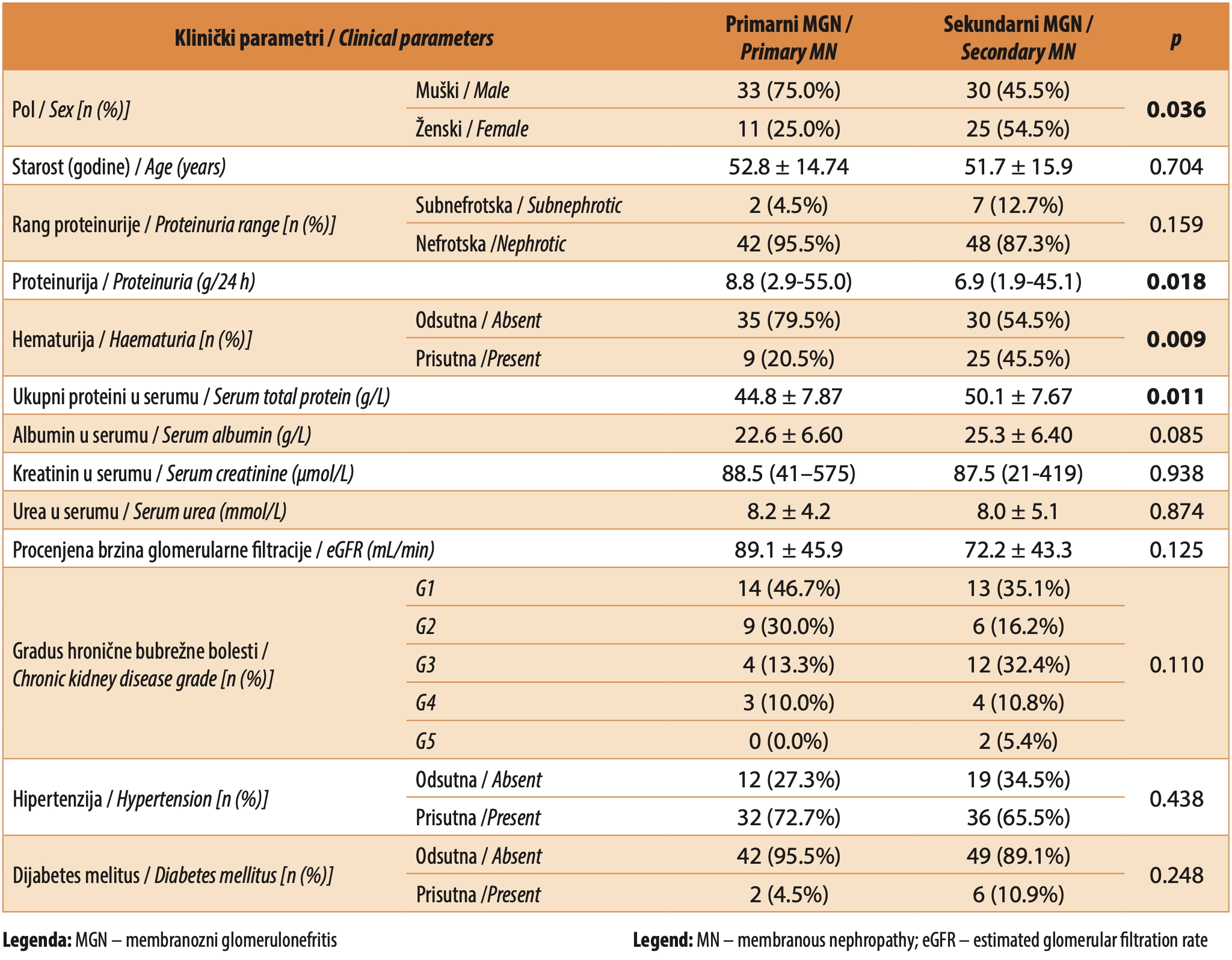
Most patients exhibited nephrotic-range proteinuria at the time of biopsy. Proteinuria was subnephrotic in 9.1% (n = 9) of cases, the majority of whom (n = 7) had secondary MGN. The median 24-hour urine protein level in patients with primary MGN (median 8.8 g/24 h; range 2.9-55.0 g/24 h) was significantly higher than in those with secondary MGN (median 6.9 g/24 h; range 1.9-45.1 g/24 h; p = 0.018). Hematuria, mostly microscopic, was observed in 34.3% (n = 34) of patients, with 73.5% (n = 25) of these cases occurring in patients with secondary MGN (p = 0.009).
The mean total protein level in patients with primary MGN was 44.8 ± 7.9 g/L, representing a significantly greater reduction (p = 0.011) compared to patients with secondary MGN (50.1 ± 7.7 g/L). Although serum albumin levels were also reduced, no significant difference was observed between the two groups (Table 1).
Indicators of excretory renal function, including serum urea and creatinine levels, estimated glomerular filtration rate, and chronic kidney disease stage, did not differ significantly between patients with primary and secondary MGN, although higher stages of chronic kidney disease (G3, G4, and G5) were more frequently observed in patients with secondary MGN (Table 1).
The most frequent comorbidities in the study population were hypertension and diabetes mellitus. Among patients with hypertension, the distribution was nearly even, with 47.1% (n = 32) having primary MGN and 52.9% (n = 36) having secondary MGN (p = 0.438). Diabetes mellitus was present in only 8.1% (n = 8) of patients, yet 75.0% (n = 6) of these cases were associated with secondary MGN (p = 0.248).
Histopathological parameters
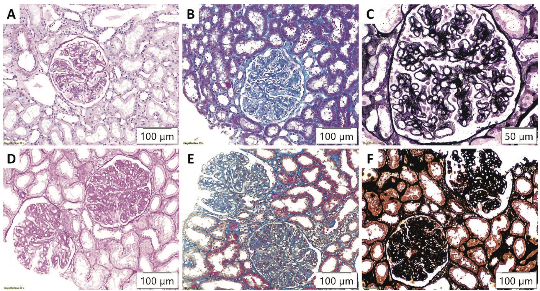
On light microscopy, all patients exhibited uniform thickening of the glomerular basement membrane, a hallmark of MGN regardless of etiology (Figure 1). Indicators of chronic glomerular damage, such as the proportion of globally and segmentally sclerotic glomeruli, did not differ significantly between primary and secondary MGN (Table 2). Crescentic glomerular formations were more common in patients with secondary MGN, although this difference was not statistically significant (p = 0.091). Endocapillary proliferation was noted in 9.1% (n = 9) of the examined tissue samples, of which 88.9% (n = 8) were from patients with secondary MGN (p = 0.032) (Table 2, Figure 1D). Karyorrhexis was observed in only two patients with secondary MGN (p = 0.500).
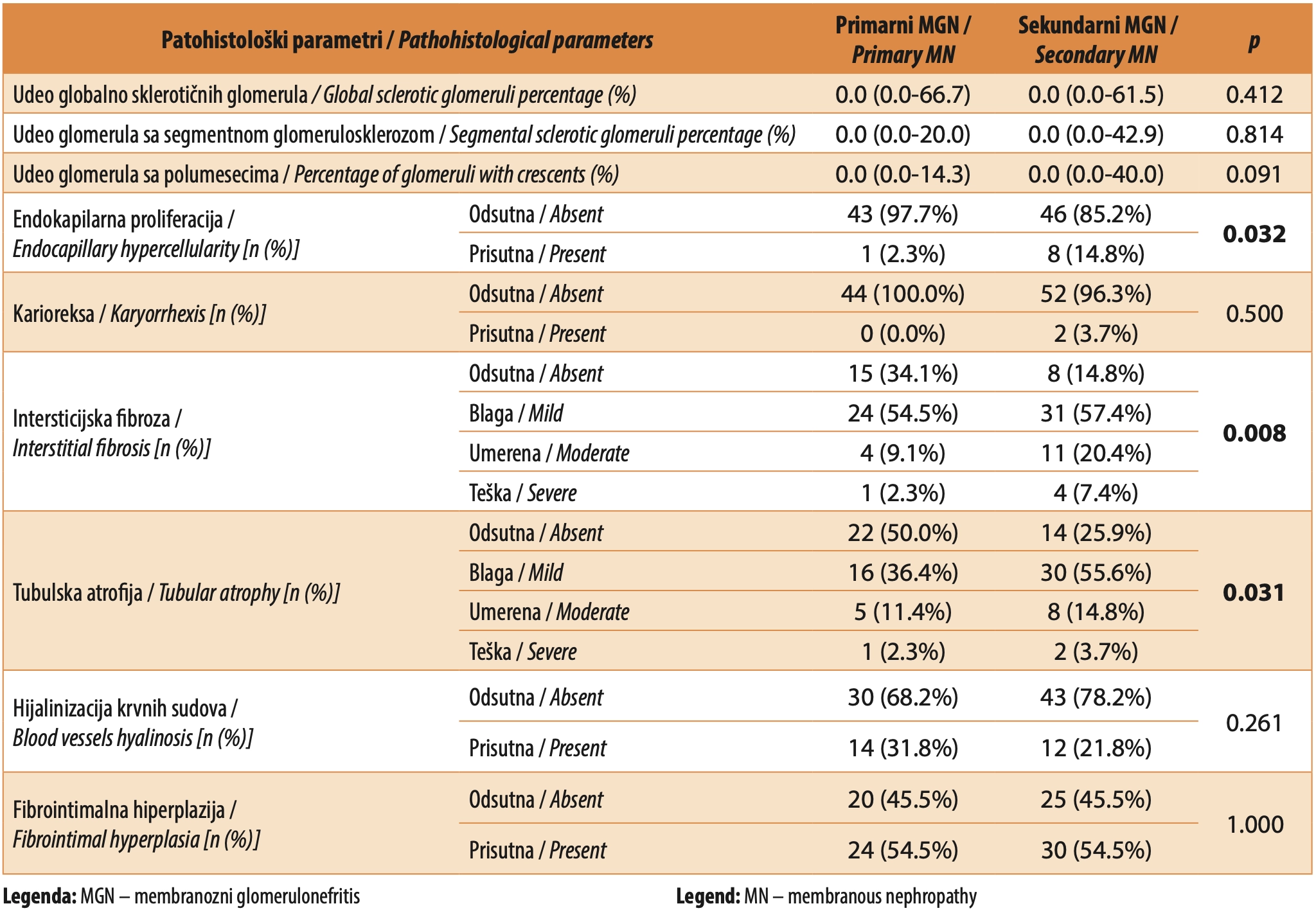
Unlike chronic vascular lesions (including medial hypertrophy, intimal fibroelastosis, and vascular hyalinization), indicators of chronic tubulointerstitial damage, such as interstitial fibrosis and tubular atrophy, differed significantly between the groups, occurring more frequently in patients with secondary MGN (Table 2).
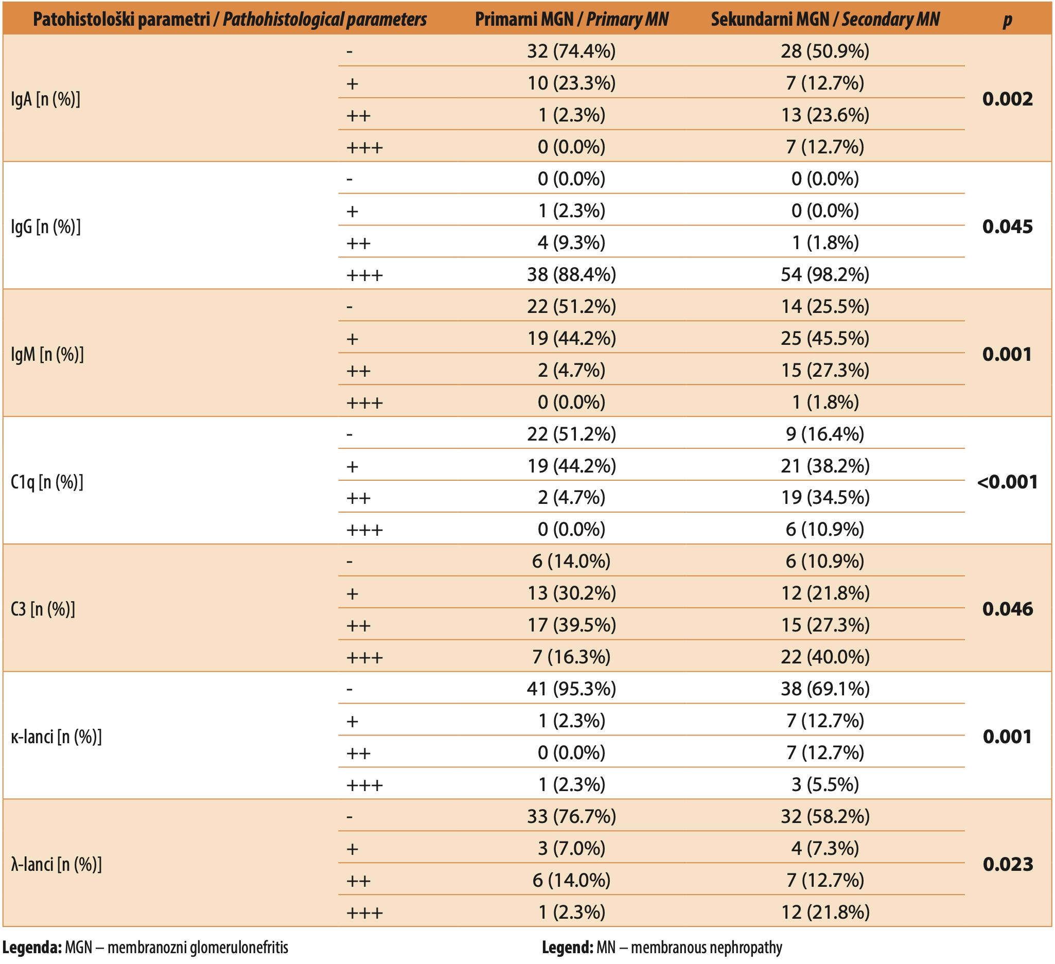
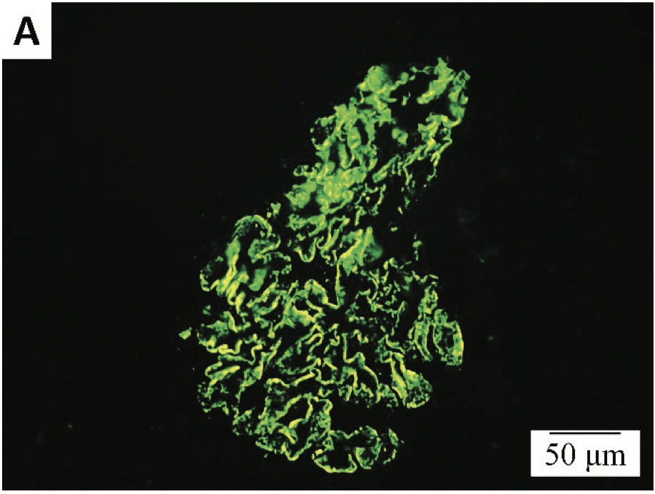
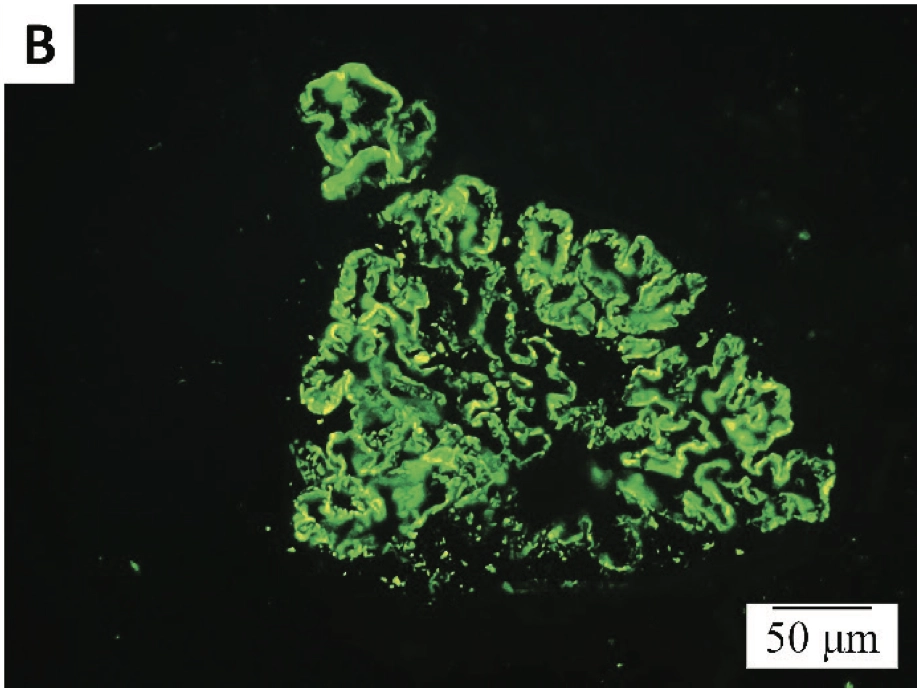
On immunofluorescence microscopy, in addition to IgG and the complement component C3, whose fine granular deposits along the glomerular basement membrane represent a characteristic finding in MGN (Figure 2), pronounced positivity for other markers (IgA, IgM, C1q, κ chains, and λ chains) was significantly more frequently observed in secondary MGN (Table 3).
DISCUSSION
In this retrospective study, clinical and pathohistological parameters were compared between patients with primary and secondary MGN. Male patients accounted for 63.6% of the study population, which is consistent with published data indicating a male-to-female ratio of approximately 1.5-2:1 in the incidence of MGN [14, 15]. Moreover, as many as 69.4% of female patients had a secondary form of the disease. This gender-based distribution may be explained by the substantial contribution of autoimmune diseases to the etiology of secondary MGN, as women are considerably more likely to develop autoimmune disorders, which can underlie the secondary form of MGN [7],[16]. The autoimmune disease most frequently associated with this pattern is systemic lupus erythematosus, in which renal involvement may manifest as membranous glomerulonephritis, corresponding to class V lupus nephritis [17]. Systemic lupus erythematosus occurs 2 to 15 times more frequently in females [18],[19].
Membranous glomerulonephritis most commonly presents with nephrotic syndrome, characterized by marked proteinuria, hypoalbuminemia, peripheral edema or anasarca, hyperlipidemia, and lipiduria [20]. Approximately one third of patients with primary MGN achieve spontaneous remission after several months or years. [21]. Proteinuria levels are an important factor in disease progression – values < 4 g/24 h are associated with a low risk, whereas values > 8 g/24 h are associated with a high risk of progression [22]. Our results demonstrated significantly higher 24-hour urinary protein levels in patients with primary MGN. Consequently, owing to greater urinary protein loss, patients with the primary form of the disease exhibited significantly reduced serum total protein levels compared with physiological values.
Literature data indicate that the presence of microscopic hematuria, arterial hypertension, and impaired renal function increases the likelihood of a secondary etiology of MGN [23],[24]. The results of our study showed that microscopic hematuria was significantly more common in patients with secondary MGN (73.5%) compared to those with primary MGN. Additionally, patients with secondary MGN had lower estimated glomerular filtration rates and a higher frequency of advanced chronic kidney disease stages (G3, G4, and G5) than those with primary MGN, although these differences were not statistically significant. In contrast, the prevalence of arterial hypertension did not differ between the groups.
In addition to serving as the gold standard for diagnosing MGN, histopathological evaluation of kidney biopsy samples can also suggest a possible etiology, whether primary or secondary. Specifically, the presence of mesangial and endocapillary hypercellularity on light microscopy is more indicative of a secondary etiology [23]. The results of our study support this conclusion–of the nine tissue samples exhibiting endocapillary proliferation, 88.9% were from patients with secondary MGN. Conversely, positivity for PLA2R and THSD7A antigens indicates a primary etiology [25],[26]. On the other hand, positivity for EXT1 and EXT2 antigens is linked to autoimmune diseases as the underlying cause of secondary MGN, primarily systemic lupus erythematosus, whereas NELL-1 positivity is associated with malignancy in approximately one-third of cases [27],[28].
The “full house” pattern on immunofluorescence microscopy, characterized by positivity for all five markers, i.e., IgG, C3, as well as IgA, IgM, and C1q, is primarily indicative of secondary MGN in the context of systemic lupus erythematosus [29]. Deposits of IgG and the complement component C3 along the glomerular basement membrane are a characteristic feature of membranous glomerulonephritis, regardless of disease etiology; however, marked positivity for these markers was more frequently observed in secondary MGN. The distribution of IgA, IgM, and C1q positivity differed significantly according to MGN etiology, with moderate to strong positivity for these markers being significantly more common in secondary MGN. Differences in κ-chain and λ-chain positivity could not be adequately interpreted, as these markers were not routinely assessed at the Institute of Pathology, Faculty of Medicine in Belgrade, until 2018.
CONCLUSION
Females are more likely to develop secondary MGN. Features of secondary MGN include hematuria, endocapillary hypercellularity, and immunofluorescence positivity for markers not typically seen in primary MGN (IgA, IgM, C1q). In contrast, primary MGN is characterized by significantly higher levels of proteinuria, accompanied by markedly reduced serum protein concentrations.
ABBREVIATION LIST
MGN – membranous glomerulonephritis
pMGN – primary membranous glomerulonephritis
sMGN – secondary membranous glomerulonephritis
Informations

-
Keywords:
-
Received:
-
Revised:
-
Accepted:
-
Online first:
-
DOI:
-
Cite this article:

Maja Životić
Institute of Pathology, Faculty of Medicine, University of Belgrade, Serbia
1 Dr Subotića starijeg Street, 11129 Belgrade, Serbia
E-mail:
1. Lai WL, Yeh TH, Chen PM, Chan CK, Chiang WC, Chen YM, et al. Membranous nephropathy: a review on the pathogenesis, diagnosis, and treatment. J Formos Med Assoc. 2015 Feb; 114(2):102-11. doi: 10.1016/j.jfma.2014.11.002. [CROSSREF]
2. Ronco P, Beck L, Debiec H, Fervenza FC, Hou FF, Jha V, et al. Membranous nephropathy. Nat Rev Dis Primers. 2021 Sep 30; 7(1):69. doi: 10.1038/s41572- 021-00303-z. [CROSSREF]
3. Menon S, Valentini RP. Membranous nephropathy in children: clinical presentation and therapeutic approach. Pediatr Nephrol. 2010 Aug; 25(8):1419- 28. doi: 10.1007/s00467-009-1324-5. [CROSSREF]
4. Glassock RJ. Membranous Nephropathy: Classification Redux? Mayo Clin Proc. 2021 Mar; 96(3):523-525. doi: 10.1016/j.mayocp.2021.01.011. [CROSSREF]
5. Beck LH Jr, Bonegio RG, Lambeau G, Beck DM, Powell DW, Cummins TD, et al. M-type phospholipase A2 receptor as target antigen in idiopathic membranous nephropathy. N Engl J Med. 2009 Jul 2; 361(1):11-21. doi: 10.1056/ NEJMoa0810457. [CROSSREF]
6. Tomas NM, Beck LH Jr, Meyer-Schwesinger C, Seitz-Polski B, Ma H, Zahner G, et al. Thrombospondin type-1 domain-containing 7A in idiopathic membranous nephropathy. N Engl J Med. 2014 Dec 11; 371(24):2277-2287. doi: 10.1056/NEJMoa1409354. [CROSSREF]
7. Moroni G, Ponticelli C. Secondary Membranous Nephropathy. A Narrative Review. Front Med (Lausanne). 2020 Dec 3; 7:611317. doi: 10.3389/ fmed.2020.611317. [CROSSREF]
8. Sethi S, Debiec H, Madden B, Charlesworth MC, Morelle J, Gross L, et al. Neural epidermal growth factor-like 1 protein (NELL-1) associated membranous nephropathy. Kidney Int. 2020 Jan; 97(1):163-174. doi: 10.1016/j. kint.2019.09.014. [CROSSREF]
9. McDonnell T, Wu HHL, Sinha S, Chinnadurai R. The Role of PLA2R in Primary Membranous Nephropathy: Do We Still Need a Kidney Biopsy? Genes (Basel). 2023 Jun 26; 14(7):1343. doi: 10.3390/genes14071343. [CROSSREF]
10. Fogo AB, Lusco MA, Najafian B, Alpers CE. AJKD Atlas of Renal Pathology: Membranous Nephropathy. Am J Kidney Dis. 2015 Sep; 66(3):e15-7. doi: 10.1053/j.ajkd.2015.07.006. [CROSSREF]
11. Kidney Disease: Improving Global Outcomes (KDIGO) Glomerular Diseases Work Group. KDIGO 2021 Clinical Practice Guideline for the Management of Glomerular Diseases. Kidney Int. 2021 Oct; 100(4S):S1-S276. doi: 10.1016/j. kint.2021.05.021. [CROSSREF]
12. Jeon SJ, Kim JH, Noh HW, Lee GY, Lim JH, Jung HY, et al. Treatment of rituximab in patients with idiopathic membranous nephropathy: a case series and literature review. Korean J Intern Med. 2022 Jul; 37(4):830-840. doi: 10.3904/kjim.2021.155. [CROSSREF]
13. Dietzen DJ, Willrich MAV. Amino Acids, Peptides, and Proteins. In: Rifai N, Chiu RWK, Young I, Burnham CAD, Wittwer CT, editors. Tietz Textbook of Laboratory Medicine. Seventh Edition. St. Louis, Missouri, USA: Elsevier; 2023. p.349.e1–349.e42.
14. Choi JY, Chin HJ, Lee H, Bae EH, Chang TI, Lim JH, et al. Idiopathic membranous nephropathy in older patients: Clinical features and outcomes. PLoS One. 2020 Oct 9; 15(10):e0240566. doi: 10.1371/journal.pone.0240566. [CROSSREF]
15. Couser WG. Primary Membranous Nephropathy. Clin J Am Soc Nephrol. 2017 Jun 7; 12(6):983-997. doi: 10.2215/CJN.11761116. [CROSSREF]
16. Ortona E, Pierdominici M, Maselli A, Veroni C, Aloisi F, Shoenfeld Y. Sex-based differences in autoimmune diseases. Ann Ist Super Sanita. 2016 Apr-Jun; 52(2):205-12. doi: 10.4415/ANN_16_02_12.
17. Almaani S, Parikh SV. Membranous Lupus Nephritis: A Clinical Review. Adv Chronic Kidney Dis. 2019 Sep; 26(5):393-403. doi: 10.1053/j.ackd.2019.08.009. [CROSSREF]
18. Pons-Estel GJ, Ugarte-Gil MF, Alarcón GS. Epidemiology of systemic lupus erythematosus. Expert Rev Clin Immunol. 2017 Aug; 13(8):799-814. doi: 10.1080/1744666X.2017.1327352. [CROSSREF]
19. Rees F, Doherty M, Grainge MJ, Lanyon P, Zhang W. The worldwide incidence and prevalence of systemic lupus erythematosus: a systematic review of epidemiological studies. Rheumatology (Oxford). 2017 Nov 1; 56(11):1945-1961. doi: 10.1093/rheumatology/kex260. [CROSSREF]
20. Alsharhan L, Beck LH Jr. Membranous Nephropathy: Core Curriculum 2021. Am J Kidney Dis. 2021 Mar; 77(3):440-453. doi: 10.1053/j.ajkd.2020.10.009. [CROSSREF]
21. Polanco N, Gutiérrez E, Rivera F, Castellanos I, Baltar J, Lorenzo D, et al. Spontaneous remission of nephrotic syndrome in membranous nephropathy with chronic renal impairment. Nephrol Dial Transplant. 2012 Jan; 27(1):231-4. doi: 10.1093/ndt/gfr285. [CROSSREF]
22. Polanco N, Gutiérrez E, Covarsí A, Ariza F, Carreño A, Vigil A, et al. Spontaneous remission of nephrotic syndrome in idiopathic membranous nephropathy. J Am Soc Nephrol. 2010 Apr; 21(4):697-704. doi: 10.1681/ASN.2009080861. [CROSSREF]
23. Basu K, Sengupta M, Mukherjee S, Karmakar S, Roychowdhury A, Bandopadhyay M. Role of light and immunofluorescence microscopy to differentiate primary and secondary membranous nephropathy. Indian J Pathol Microbiol. 2022 Oct-Dec; 65(4):821-827. doi: 10.4103/ijpm.ijpm_22_21. [CROSSREF]
24. Dantas M, Silva LBB, Pontes BTM, Dos Reis MA, de Lima PSN, Moysés Neto M. Membranous nephropathy. J Bras Nefrol. 2023 Apr-Jun; 45(2):229-243. doi: 10.1590/2175-8239-JBN-2023-0046en. [CROSSREF]
25. Larsen CP, Messias NC, Silva FG, Messias E, Walker PD. Determination of primary versus secondary membranous glomerulopathy utilizing phospholipase A2 receptor staining in renal biopsies. Mod Pathol. 2013 May; 26(5):709- 15. doi: 10.1038/modpathol.2012.207. [CROSSREF]
26. Lwezaula BF, Ameh OI, Ekrikpo UE, Botha FC, Okpechi-Samuel US, Wearne N, et al. Diagnostic performance of glomerular PLA2R and THSD7A antibodies in biopsy confirmed primary membranous nephropathy in South Africans. BMC Nephrol. 2021 Jan 7; 22(1):15. doi: 10.1186/s12882-020-02216-7. [CROSSREF]
27. Sethi S, Madden BJ, Debiec H, Charlesworth MC, Gross L, Ravindran A, et al. Exostosin 1/Exostosin 2-Associated Membranous Nephropathy. J Am Soc Nephrol. 2019 Jun; 30(6):1123-1136. doi: 10.1681/ASN.2018080852. [CROSSREF]
28. Caza TN, Hassen SI, Dvanajscak Z, Kuperman M, Edmondson R, Herzog C, et al. NELL1 is a target antigen in malignancy-associated membranous nephropathy. Kidney Int. 2021 Apr; 99(4):967-976. doi: 10.1016/j.kint.2020.07.039. [CROSSREF]
29. Cameron JS. Lupus nephritis. J Am Soc Nephrol. 1999 Feb; 10(2):413-24. doi: 10.1681/ASN.V102413. [CROSSREF]
- Table 1. Clinical parameters of subjects with membranous nephropathy
- Figure 1. Changes in glomeruli on light microscopy in primary (A) Periodic acidSchiff (PAS), x200, (B) Masson’s trichrome (MTS), x200, (C) Jones methenamine silver (JMS), x400; and secondary membranous nephropathy, with evident endocapillary hypercellularity (D) PAS, x200, (E) MTS, x200, (F) JMS, x200
- Table 2. Pathohistological parameters of patients with membranous nephropathy by light microscopy
- Table 3. Pathohistological parameters of patients with membranous nephropathy by immunofluorescence
- Figure 2. Immunofluorescence microscopy findings: fine granular deposits of (A) IgG and (B) C3 along the glomerular basement membrane, x400
- Figure 2. Immunofluorescence microscopy findings: fine granular deposits of (A) IgG and (B) C3 along the glomerular basement membrane, x400