


Prognostic and predictive significance of measurable residual disease in acute myeloblastic leukemia
- University Clinical Center of Serbia, Clinic for Hematology, Belgrade, Serbia
- University of Belgrade, Faculty of Medicine, Belgrade, Serbia
ABSTRACT
Introduction: Acute myeloblastic leukemia (AML) is an umbrella term for a heterogeneous group of clonal neoplastic diseases of hematopoietic cells. Detecting residual leukemic cells (measurable residual disease – MRD) is the most important prognostic and predictive factor in AML.
The aim: The study aims to analyze the effect of administered chemotherapy based on the results of MRD testing in patients with AML treated at the University Clinical Center of Serbia (UCCS) Clinic for Hematology.
Materials and methods: Our study included the analysis of 111 AML patients, treated between January 2020 and January 2024. All diagnostic procedures performed were based on the most recent recommendations of European LeukemiaNet (ELN).
Results: MRD+ patients who continued treatment with intensive chemotherapy (CHT), using full doses of 3+7 CHT as reinduction therapy, had a significantly longer remission (complete remission – CR) and a longer overall survival (OS). The duration of CR (p = 0.004) and OS (p = 0.019) were statistically significantly longer in patients who maintained a negative MRD status at the end of treatment. In transplanted patients, overall survival (OS; p = 0.006) and duration of remission (CR; p = 0.002) were significantly longer (median: OS 20 months; CR 21 months), as compared to the group of non-transplanted patients (median: OS 13 months; CR 8 months).
Discussion: Measurable residual disease (MRD) can be both prognostic and predictive. However, the absolute measurable level of the disease is not the only determinant of the patient’s outcome, since the biology of AML, as well as other clinical patient-related factors (age, comorbidities, various complications of applied chemotherapy, especially infections), modify the risk associated with MRD test results.
Conclusion: The study has demonstrated the great importance of timely detection of MRD, as well as the appropriateness of applying more intensive CHT in MRD-positive patients, along with continued treatment with allogeneic hematopoietic stem cell transplantation.
INTRODUCTION
Acute myeloblastic leukemia (AML) is an umbrella term for a heterogeneous group of clonal neoplastic diseases of hematopoietic cells. The use of intensive chemotherapy, with or without the use of small molecule inhibitors or immunotherapy, leads to a high percentage of cytomorphological remission of about 80%, especially in patients younger than 60 years. The problem is the high rate of relapse, which is 30% – 60% within two years, which represents the main barrier to achieving complete cure in AML [1]. It has long been known that cytomorphological remission, which has a sensitivity level of about 1%, i.e. the presence of less than 5% blasts, counted in 300 to 500 nucleated cells, is not a guarantee of complete cure when it comes to AML [1],[2].
Detecting residual leukemic cells, originally referred to as minimal residual disease (MRD) and subsequently measurable residual disease (MRD), is the most important prognostic and predictive factor in AML [3].
In the last five years, meta-analyses have shown that, regardless of the methodology used to detect MRD, patients who were positive, either at a certain point in time before allogeneic hematopoietic stem cell transplantation (alloTMHC) or in general, during any period of treatment for acute myeloblastic leukemia, have significantly shorter survival compared to MRD negative patients [4],[5].
There are three main techniques for detecting MRD in AML: multiparametric flow-cytometry (MFC), quantitative polymerase chain reaction (qPCR), and next-generation sequencing (NGS) [1],[4],[5].
Immunophenotyping by MFC is crucial for the diagnosis of acute myeloblastic leukemia, but also for MRD detection. Precisely applied markers achieve a sensitivity of 10-3. There are two approaches to this method. The first approach is based on the identification of one or more immunophenotypes associated with leukemia (leukemia-associated immunophenotype – LAIP) at diagnosis and monitoring throughout the entire treatment. The second approach focuses on the detection of cell populations deviating from antigen expression patterns characteristic of normal or regenerating cells, the so-called difference from normal (DfN). A combination of both approaches is recommended [6],[7].
The polymerase chain reaction (PCR) method has a sensitivity of 10-4 to 10-5. This method can detect DNA mutations at very sensitive levels. Polymerase chain reaction represents a more sensitive and standardized way of quantifying a specific mutation, which seems to be an ideal method for the detection of low levels of residual leukemic cells. However, this method also has several limitations. Perhaps the most important limitation is the heterogeneity of acute myeloblastic leukemia itself since, despite numerous cytogenetic and molecular mutations, the genome is relatively stable in AML. Exceptions are late stages of AML, as well as the presence of a TP53 mutation or a complex karyotype [8],[9].
Next-generation DNA and RNA sequencing is the most sensitive technique (up to 10-7 sensitivity), but standards for this technique are still being established. The main limitation of this technique, when it comes to detecting MRD, is the fact that AML is a “tornado” of low mutational status, where mutations are often not in one point, but gene fusions or other structural variations, which are more difficult to detect with the current NGS approach. Also, another limitation of NGS is that variants with a frequency of less than 1% cannot be detected, due to the error rate of conventional NGS, thus opening the possibility of a false negative result [10],[11],[12].
After 60 years of relative inactivity in updating response criteria in AML, in 2017, the European Leukemia Network (ELN) introduced a new category of MRD-negative complete remissions [13]. In 2018, this was followed by the announcement of the first ELN consensus on standard treatment guidelines, which contains detailed instructions for the application of flow cytometry, molecular testing, and clinical interpretations of MRD in AML [14].
The ELN AML MRD guidelines have been updated (with the expectation that these evidence-based guidelines will be updated every two to four years). They contain almost 60 different recommendations covering a range of MRD detection methods, such as MRD by multiparametric flow cytometry and molecular MRD detection, as well as guidelines for the clinical use of MRD and instructions for future developments in the field of MRD [15].
STUDY AIM
Aware of the great prognostic and predictive significance of measurable residual disease, we have analyzed the significance of MRD in patients with AML treated at the University Clinical Center of Serbia (UCCS) Clinic for Hematology, with the aim of presenting the MRD testing strategy, the modalities of the therapy applied, especially in MRD-positive (MRD+) patients, and the survival of patients with AML in relation to the MRD values.
MATERIALS AND METHODS
The diagnosis was established using a myelogram. The immunophenotypic characteristics of AML were determined at diagnosis; cytogenetic/molecular testing was performed; risk stratification of AML patients was carried out based on ELN criteria [16]. To determine the MRD status, the method of multiparametric flow cytometry was applied, using an eight-color flow cytometer [17],[18].
Statistical analysis
Depending on the type of variables and the normality of distribution, data is presented as n (%) or median (min-max). The Kaplan-Meier method was used for survival analysis. The log-rank test was used to evaluate the survival function of these patients depending on transplantation and positive MRD after three or more cycles of chemotherapy, and the Cox proportional regression model with a 95% confidence interval was used to evaluate the independent predictor – fatal outcome. Data were censored for the following reasons: the subject survived the entire follow-up period or was lost from the records. Statistical hypotheses were tested at a statistical significance level (alpha level) of 0.05. All data were processed with the IBM SPSS Statistics 24 (SPSS Inc., Chicago, IL, USA) software package or in the R programming environment (R Core Team, 2023).
RESULTS
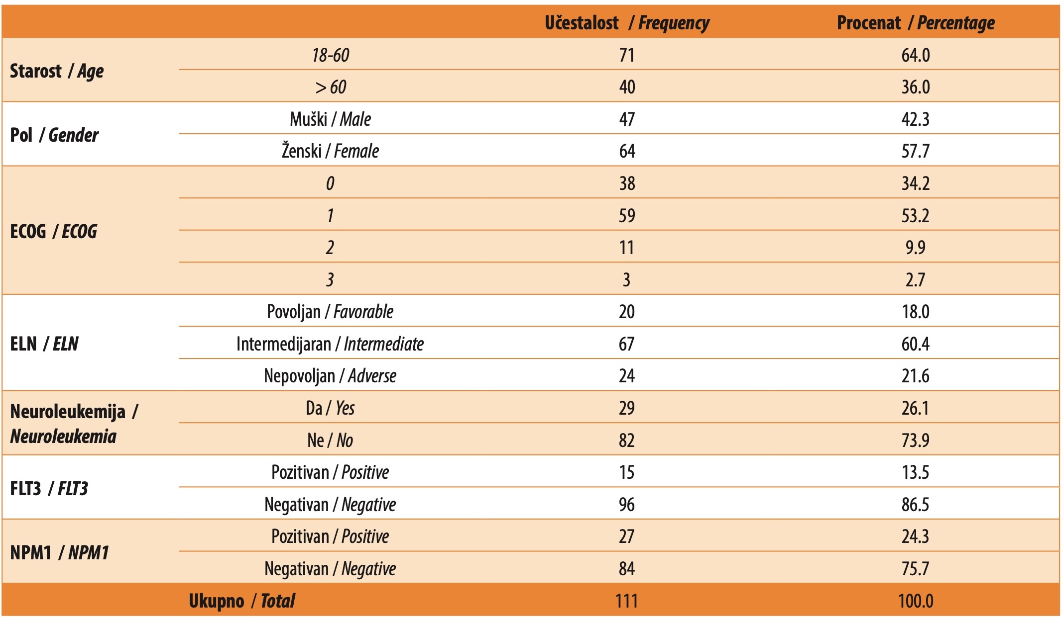
Our study analyzed 111 AML patients, treated at the UCCS Clinic for Hematology, between January 2020 to January 2024. All patients received at least two or more cycles of chemotherapy (CHT). MRD assessment was performed after two cycles of chemotherapy (induction and reinduction or induction and first consolidation) and at the end of chemotherapy treatment. The median age of the examined patients was 55 years (18-75 years). Out of 111 patients, 71 (64%) patients were aged 18-60 years, while 40 (36%) patients were older than 60 years. Out of the total number, 64 (57.7%) patients were female, while 47 (42.3%) were male. The largest number of patients, 59 (53.2%) in total, had ECOG performance status (PS) 1. Neuroleukemia was present in 29 (26.1%) patients. According to the ELN classification, most patients were classified in the intermediate risk group, i.e. 67 (60.4%) patients, 20 (18.0%) patients were in the favorable risk group, and 24 (21.6%) patients were in the adverse risk group. FMS-like tyrosine kinase 3 internal tandem duplication (FLT3-ITD) positivity was present in 15 (13.5%) patients, while the nucleophosmin 1 (NPM1) mutation was positive in 27 (24.3%) patients. These results are presented in Table 1.
The 3+7 regimen was the most frequently applied induction chemotherapy regimen. It was used in 61 (55%) patients. Seven (6.3%) patients received the FMS-like tyrosine kinase 3 (FLT3) inhibitor (midostaurin) in addition to the 3+7 regimen, while 25 (22.5%) patients received the 3+7 light regimen, and 18 (16.2%) patients received the 2+5 regimen.
Cytomorphological remission after induction CHT was achieved by 64 (57.7%) patients. Cytomorphological remission after reinduction CHT was achieved in another 20 (18%) patients, while the remaining 27 (24.3%) patients were refractory to the applied CHT. Thus, after induction and reinduction, there were 84 (75.7%) patients in cytomorphological remission.
After two cycles of CHT were administered, MRD negativity was achieved in 35 (31.5%) patients, who were all subsequently treated with IDAC consolidation therapy. Seventy-six (68.5%) patients who were treated with various CHT protocols had a positive MRD value: 31 (40.8%) patients received Mitox-VP, 19 (25%) patients received IDAC+DA, IDA-VP was administered to 8 (10.5%) patients, MEC was received by 7 (9.2%) patients, 7 (9.2%) patients received the 3+7 regimen, IDAC was given to three (4.0%) patients, while FLAG IDA was received by one (1.3%) patient (Graph 1).

In 81 (73%) patients, MRD testing was also performed at the end of treatment. Of these patients, 46 were MRD-negative, while 35 patients were MRD-positive. Also, out of 50 patients who demonstrated MRD positivity after the second cycle of CHT, 28 (56%) patients achieved MRD negativity at the end of treatment. Disease relapse occurred in 43 (37.8%) patients.
Median overall survival (OS) in our patients was 13 months (2-69). The median length of cytomorphological remission in our patients was 9 months (0-64). Median survival in younger patients (18-60 years) was 18.0 months (95% CI = 13.3-22.7), while in older (> 60 years) patients it was 13.0 months (95% CI = 7.5-18.5), which is a statistically significant difference (log-rank = 3.722; p = 0.054). The length of remission (complete remission – CR) was significantly longer in the age group 18-60 years, as compared to the group > 60 years (p = 0.02, HR = 0.568, 95% CI = 0.352-0.915).
There was no statistically significant difference in overall survival (OS) and length of remission (CR) in relation to gender (p = 0.245, HR = 1.321, 95% CI = 0.826-2.114). No statistically significant difference was found in overall survival (OS) and length of CR in relation to MRD I status (after the first consolidation or after two cycles of initial reinduction therapy), (p = 0.380; p = 0.288).
However, median survival in patients with a positive MRD test at the end of treatment was 16.0 months (95% CI = 9.3-22.7), while in MRD-negative patients it was 36.0 months (95% CI = 17.6-54.4), which is a statistically significant difference (p = 0.019). Subjects with positive MRD at the end of treatment had more than twice the hazard (HR = 2.06, 95% CI = 1.10-3.85; p = 0.024), (Graph 2). Also, the length of CR was statistically significantly longer in patients with negative MRD at the end of treatment (p = 0.004, HR = 2.517, 95% CI = 1.342-4.720).

Overall survival (OS) was significantly longer in the group with intermediate risk of leukemia (median 18 months) according to the ELN classification, compared to the group with favorable (median 13 months) and adverse (median 6 months) risk (p = 0.013). Also, the duration of remission (CR) was significantly longer in the group of patients with intermediate risk (median 14 months), compared to favorable (median 11 months) and adverse risk (median 5 months), (p = 0.014, HR = 0.503, 95% CI = 0.291-0.871).
Overall survival (OS) was significantly longer in patients in whom reinduction with full doses of the 3+7 regimen was applied after a positive MRD test (median survival 35 months), while survival was significantly shorter with the application of other protocols: IDAC (median 15.5 months), IDAC+DA (median 13 months), MEC (median 10 months), Mitox-VP (median 10 months), IDA-VP (median 7.5 months).
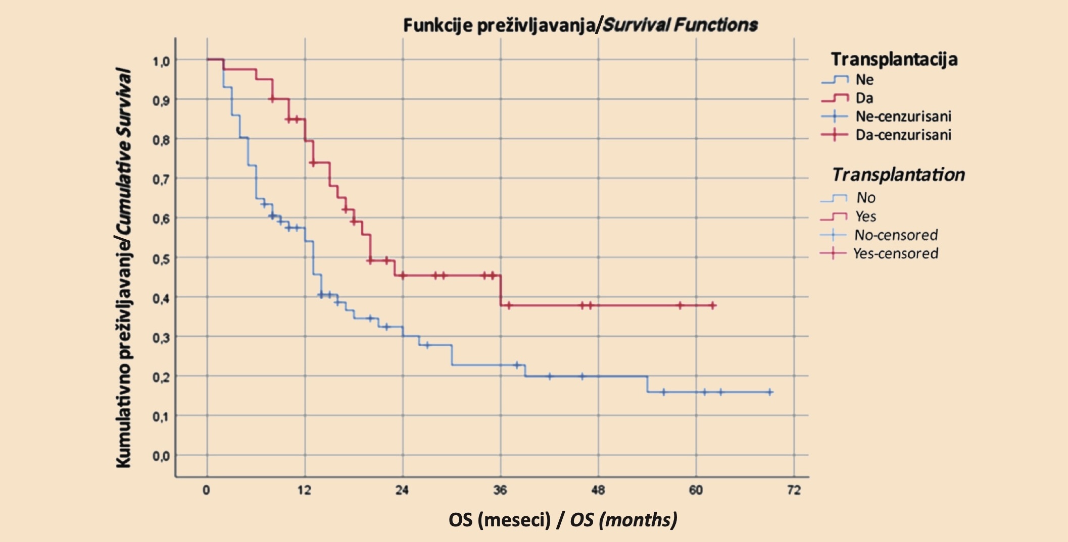
Transplantation was performed in 40 patients (36%). The indications for transplantation were as follows: adverse risk (ELN), found in six (15%) patients, disease relapse, found in 12 (30%) patients, and a positive MRD test, which was found in 22 (55%) patients. Median survival in transplanted patients was 20 months (95% CI = 4.0-36.0), while in non-transplanted patients it was 13 months (95% CI = 10.1-15.9), which is a statistically significant difference (p = 0.006). Transplant subjects had a 50% lower risk of death (HR = 0.498, 95% CI = 0.296-0.839; p = 0.009), (Chart 3). The duration of remission (CR) was significantly longer (21 months) in transplanted patients, compared to the group of non-transplanted patients (8 months), (p = 0.002, HR = 0.441, 95% CI = 0.261-0.745).
DISCUSSION
As a biomarker, measurable residual disease (MRD) can be both prognostic and predictive. However, the absolute measurable level of the disease is not the only determinant of the patient’s outcome, since the biology of AML, as well as other clinical patient-related factors (age, comorbidities, various complications of applied chemotherapy, especially infections), modify the risk associated with MRD test results [19],[20],[21].
Clearly, not all mutations detected at AML diagnosis will be equally clinically useful for MRD monitoring. Optimal targets for molecular MRD measurement have also not been defined.
Isolated detection of mutations common in age-related clonal hematopoiesis, e.g. DNMT3A, TET2, or ASKSL1, or in germline predisposition syndromes, e.g. DDKS41, RUNKS1, or GATA2, do not necessarily constitute residual AML [17],[18],[19],[20].
Our study has demonstrated no significant difference in CR and OS length among patients, with regard to MRD status after two cycles of CHT. On the other hand, the type of CHT used in MRD+ patients had a significant impact on the length of CR, as well as on OS, with significantly longer CR and OS in patients who, after MRD+, continued treatment with full doses of 3+7 CHT, as reinduction therapy. Also, CR and OS were statistically significantly longer in patients who maintained a negative MRD status at the end of treatment.
In relation to the ELN risk stratification, patients in the intermediate-risk group had the longest OS. One of the possible explanations is that the patients from the favorable-risk group, who were expected to have the longest OS, had certain gene mutations that we did not detect since, for now, testing for these mutations is not part of the standard tests performed at our clinic.
Patients treated with allogeneic HSCT had a statistically longer duration of CR and OS, as compared to non-transplanted patients, as demonstrated in several large studies [22],[23],[24].
CONCLUSION
Analysis of the results of this study, as well as of the data from literature, has yielded several conclusions.
Existing data indicate that mutations in signaling pathway genes (FLT3, KIT, RAS, etc.) are useful when they are positive, but their negativity in later follow-up does not necessarily mean molecular remission, especially when the mutation is therapeutically targeted with the appropriate agents. Also, it is debatable whether MRD negativity has the same prognostic significance if it is achieved after intensive versus non-intensive therapy.
As a biomarker, measurable residual disease (MRD) can have both prognostic and predictive value. However, the absolute measurable level of disease is not the only determinant of patient outcome, as AML biology and other clinical factors modify the risk associated with MRD test results.
Our study emphasizes the great importance of timely detection of MRD, as well as the appropriateness of applying more intensive CHT in MRD-positive patients, along with continued treatment with allogeneic hematopoietic stem cell transplantation.
Informations

-
Keywords:
-
Received:
-
Revised:
-
Accepted:
-
Online first:
-
DOI:
-
Cite this article:

Ana Vidović
Clinic for Hematology, University Clinical Center of Serbia
2 Dr Koste Todorovića Street, 11000 Belgrade, Serbia
E-mail:
1. Blachly JS, Walter RB, Hourigan CS. The present and future of measurable residual disease testing in acute myeloid leukemia. Haematologica. 2022 Dec 1;107(12):2810-22. doi: 10.3324/haematol.2022.282034. [CROSSREF]
2. Short NJ, Zhou S, Fu C, Berry DA, Walter RB, Freeman SD, et al. Association of measurable residual disease with survival outcomes in patients with acute myeloid leukemia: a systematic review and meta-analysis. JAMA Oncol. 2020 Dec 1;6(12):1890-9. doi: 10.1001/jamaoncol.2020.4600. [CROSSREF]
3. Heuser M, Freeman SD, Ossenkoppele GJ, Buccisano F, Hourigan CS, Ngai LL, et al. 2021 Update on MRD in acute myeloid leukemia: a consensus document from the European LeukemiaNet MRD Working Party. Blood. 2021 Dec 30;138(26):2753-67. doi: 10.1182/blood.2021013626. [CROSSREF]
4. Walter RB, Ofran Y, Wierzbowska A, Ravandi F, Hourigan CS, Ngai LL, et al. Measurable residual disease as a biomarker in acute myeloid leukemia: theoretical and practical considerations. Leukemia. 2021 Jun;35(6):1529-38. doi: 10.1038/s41375-021-01230-4. [CROSSREF]
5. Caballero-Velázquez T, Pérez-López O, Yeguas Bermejo A, Rodríguez Arbolí E, Colado Varela E, Sempere Talens A et al. Prognostic value of measurable residual disease in patients with AML undergoing HSCT: a multicenter study. Cancers (Basel). 2023 Mar 5;15(5):1609. doi: 10.3390/cancers15051609. [CROSSREF]
6. Tettero JM, Freeman S, Buecklein V, Venditti A, Maurillo L, Kern W, et al. Technical aspects of flow cytometry-based measurable residual disease quantification in acute myeloid leukemia: experience of the European LeukemiaNet MRD Working Party. Hemasphere. 2021 Dec 22;6(1):e676. doi: 10.1097/ HS9.0000000000000676. [CROSSREF]
7. Li SQ, Xu LP, Wang Y, Zhang XH, Chen H, Chen YH, et al. An LSC-based MRD assay to complement the traditional MFC method for prediction of AML relapse: a prospective study. Blood. 2022 Aug 4;140(5):516-20. doi: 10.1182/ blood.2021014604. [CROSSREF]
8. Orvain C, Wilson JA, Fang M, Sandmaier BM, Rodríguez-Arbolí E, Wood BL, et al. Relative impact of residual cytogenetic abnormalities and flow cytometric measurable residual disease on outcome after allogeneic hematopoietic cell transplantation in adult acute myeloid leukemia. Haematologica. 2023 Feb 1;108(2):420-32. doi: 10.3324/haematol.2022.281585. [CROSSREF]
9. Schuurhuis GJ, Heuser M, Freeman S, Béné MC, Buccisano F, Cloos J, et al. Minimal/measurable residual disease in AML: a consensus document from the European LeukemiaNet MRD Working Party. Blood. 2018 Mar 22;131(12):1275-91. doi: 10.1182/blood-2017-09-801498. [CROSSREF]
10. Heuser M, Heida B, Büttner K, Wienecke CP, Teich K, Funke C, et al. Posttransplantation MRD monitoring in patients with AML by next-generation sequencing using DTA and non-DTA mutations. Blood Adv. 2021 May 11;5(9):2294-304. doi: 10.1182/bloodadvances.2021004367. [CROSSREF]
11. Duncavage EJ, Schroeder MC, O’Laughlin M, Wilson R, MacMillan S, Bohannon A, et al. Genome sequencing as an alternative to cytogenetic analysis in myeloid cancers. N Engl J Med. 2021 Mar 11;384(10):924-35. doi: 10.1056/NEJMoa2024534. [CROSSREF]
12. Patkar N, Kakirde C, Shaikh AF, Salve R, Bhanshe P, Chatterjee G, et al. Clinical impact of panel-based error-corrected next generation sequencing versus flow cytometry to detect measurable residual disease (MRD) in acute myeloid leukemia (AML). Leukemia. 2021 May;35(5):1392-404. doi: 10.1038/ s41375-021-01131-6. [CROSSREF]
13. Döhner H, Estey E, Grimwade D, Amadori S, Appelbaum FR, Büchner T, et al. Diagnosis and management of AML in adults: 2017 ELN recommendations from an international expert panel. Blood. 2017 Jan 26;129(4):424-47. doi: 10.1182/blood-2016-08-733196. [CROSSREF]
14. Schuurhuis GJ, Heuser M, Freeman S, Béné MC, Buccisano F, Cloos J, et al. Minimal/measurable residual disease in AML: a consensus document from the European LeukemiaNet MRD Working Party. Blood. 2018 Mar 22;131(12):1275-91. doi: 10.1182/blood-2017-09-801498. [CROSSREF]
15. Döhner H, Pratz KW, DiNardo CD, Jonas BA, Pullarkat VA, Thirman MJ, et al. ELN risk stratification is not predictive of outcomes for treatment-naïve patients with acute myeloid leukemia treated with venetoclax and azacitidine. Blood. 2022 Nov 15;140(Suppl 1):1441-4. doi: 10.1182/blood-2022-169509. [CROSSREF]
16. Döhner H, Wei AH, Appelbaum FR, Craddock C, DiNardo CD, Dombret H, et al. Diagnosis and management of AML in adults: 2022 recommendations from an international expert panel on behalf of the ELN. Blood. 2022 Sep 22;140(12):1345-77. doi: 10.1182/blood.2022016867. [CROSSREF]
17. Moritz J, Schwab A, Reinisch A, Zebisch A, Sill H, Wölfler A. measurable residual disease detection in acute myeloid leukemia: current challenges and future directions. Biomedicines. 2024 Mar 7;12(3):599. doi: 10.3390/biomedicines12030599. [CROSSREF]
18. Döhner H, Wei AH, Löwenberg B. Towards precision medicine for AML. Nat Rev Clin Oncol. 2021 Sep;18(9):577-90. doi: 10.1038/s41571-021-00509-w. [CROSSREF]
19. Tanaka T, Morita K, Loghavi S, Wang F, Furudate K, Sasaki Y, et al. Clonal dynamics and clinical implications of postremission clonal hematopoiesis in acute myeloid leukemia. Blood. 2021 Nov 4;138(18):1733-9. doi: 10.1182/ blood.2020010483. [CROSSREF]
20. Gui G, Hourigan CS. Measurable residual disease assessment as a surrogate marker in new drug development in acute myeloid leukemia. Cancer J. 2022 Jan-Feb 01;28(1):73-7. doi: 10.1097/PPO.0000000000000572. [CROSSREF]
21. Rodríguez-Arbolí E, Othus M, Orvain C, Zarling LC, Sandmaier BM, Milano F, et al. Contribution of measurable residual disease status to prediction accuracy of relapse and survival in adults with acute myeloid leukemia undergoing allogeneic hematopoietic cell transplantation. Haematologica. 2023 Jan 1;108(1):273-7. doi: 10.3324/haematol.2022.281631. [CROSSREF]
22. Hourigan CS, Dillon LW, Gui G, Logan BR, Fei M, Ghannam J et al. Impact of Conditioning Intensity of Allogeneic Transplantation for Acute Myeloid Leukemia With Genomic Evidence of Residual Disease. J Clin Oncol. 2020 Apr 20;38(12):1273-83. doi: 10.1200/JCO.19.03011. [CROSSREF]
23. Wang T, Zhou B, Zhang J, Zhang X, Liu T, Qiu H, et al. Allogeneic hematopoietic stem cell transplantation could improve survival for pure CBF-AML patients with minimal residual disease positive after the second consolidation. Leuk Lymphoma. 2021 Apr;62(4):995-8. doi: 10.1080/10428194.2020.1846736. [CROSSREF]
24. Rautenberg C, Lauseker M, Kaivers J, Jäger P, Fischermanns C, Pechtel S, et al. Prognostic impact of pretransplant measurable residual disease assessed by peripheral blood WT1-mRNA expression in patients with AML and MDS. Eur J Haematol. 2021 Aug;107(2):283-92. doi: 10.1111/ejh.13664. [CROSSREF]
- Table 1. Initial clinical characteristics of the examined patients
- Graph 1. Type of therapy used in MRD-positive patients (after the second cycle of initial therapy) After two cycles of CHT were administered, MRD negativity was achieved in 35 (31.5%) patients and all of them were subsequently treated with consolidation IDAC therapy. Seventy-six (68.5%) patients had a positive MRD and were treated with different CHT protocols: 31 (40.8%) patients received Mitox-VP; 19 (25%) patients received IDAC+DA; 8 (10.5%) patients received IDA-VP; 7 (9.2%) patients received MEC; 3+7 was received by 7 (9.2%) patients; three (4.0%) patients received IDAC; FLAG IDA was received by one (1.3%) patient.
- Graph 2. Kaplan-Meier curve of overall survival (OS) in relation to the MRD status at the end of treatment Median survival in patients with a positive MRD at the end of treatment was 16.0 months (95% CI = 9.3-22.7), while it was 36.0 months (95% CI = 17.6-54.4) in patients with a negative MRD, which is a statistically significant difference (p = 0.019). Subjects with a positive MRD at the end of treatment had more than twice the hazard for a fatal outcome (HR = 2.06, 95% CI = 1.10-3.85; p = 0.024).
- Graph 3. Kaplan-Meier curve of overall survival (OS) in patients on chemotherapy in relation to transplant patients The median survival in transplant patients was 20 months (95% CI = 4.0-36.0), while in non-transplant patients it was 13 months (95% CI = 10.1-15.9), which is a statistically significant difference (p = 0.006). Subjects who had received a transplant had a 50% lower hazard for the fatal outcome (HR = 0.498, 95% CI = 0.296-0.839; p = 0.009).