


Light chain deposition disease
- University Clinical Center Kragujevac, Clinic for Hematology, Kragujevac, Serbia
- University of Kragujevac, Faculty of Medical Sciences, Department of Internal Medicine, Kragujevac, Serbia
ABSTRACT
Light chain deposition disease is one form of monoclonal immunoglobulin deposition disease. In terms of frequency, it is a rare entity that occurs in middle-aged people, more often males. It most often affects the kidneys, with a clinical picture of nephritic syndrome, but it can also be localized in other organs, such as the liver, lungs, heart, gastrointestinal tract, skin, and others. The symptoms and signs of the affected organ dominate the clinical picture. The diagnosis is most often established by biopsy of the affected organ, whereby Congo red staining differentiates it from light chain amyloidosis, and then bone marrow evaluation is performed to rule out other plasmacytic dyscrasias. Therapy is based on therapeutic modalities for treating multiple myeloma, including proteasome inhibitors (bortezomib), autologous hematopoietic stem cell transplantation, and transplantation of the affected organs if there has been a complete loss of function. Data on monoclonal antibody therapy (daratumumab) opens up new therapeutic possibilities for the treatment of this disease. Diagnosis and treatment of this disease require a multidisciplinary approach, primarily by nephrologists, hematologists, and pathologists.
INTRODUCTION
Light chain deposition disease, according to the 5th revision of the classification of hematopoietic and lymphoid tissue diseases of the World Health Organization, belongs, as the most common entity, to the group of monoclonal immunoglobulin deposition diseases, which also includes heavy chain deposition diseases, as well as light and heavy chain deposition diseases [1]. The deposition of immunoglobulin light chains in various organs characterizes it. Precipitated light chains do not have the characteristics of an amyloid substance, distinguishing this disease from light chain amyloidosis [2].
The disease was first proven in 1976 in the kidney biopsies of two patients in the United States of America. Both patients had kidney failure, and other manifestations developed very quickly, first liver weakness and neurological symptoms, and then heart failure and gastrointestinal and endocrine manifestations [3].
Given the recently limited possibilities for precise detection of light chains, the frequency of this disease is unknown. It occurs more often in men, and the median age at diagnosis is 58, while as many as 36% of patients are younger than 50 [4].
ETIOLOGY AND PATHOGENESIS
The etiology of this disease is unknown.
At the basis of the disease, a small pathological clone of plasma cells with a low proliferative potential produces light chains, predominantly of the kappa type, as a pathological protein [5]. Free monoclonal light chains are deposited in various organs as disorganized, amorphous aggregates without forming deposits in the form of fibrillar strands, which is the main difference at the microscopic level between light chain amyloidosis and light chain deposition disease [6]. The disease of deposition of light chains is most often manifested in the kidneys (in about 85% of patients), less often in the liver, heart, and lungs, and very rarely in other organs [4].
The pathophysiological mechanism of impaired function is best studied in renal manifestations, where amorphous aggregates of light chains initiate a cascade of events leading to mesangial expansion and enlargement of an extracellular matrix of unusual composition (rich in tenascin), forming mesangial nodules that replace the standard matrix rich in collagen IV [5],[6]. Tubulointerstitial kidney disease is also mentioned in the literature as a rarer but sometimes the first manifestation of light chain deposition disease. Most often, deposits of light chains are seen along the tubular basement membrane, along with linear glomerular and vascular deposits [7]. Rarely, aggregates of light chains and Tamm Horsfall protein can lead to distal tubule obstruction and acute renal failure [8],[9]. Although changes in renal structures can be detected by light microscopy, the gold standard of diagnosis is electron microscopy [10]. On preparations under the electron microscope, small, granular, dense deposits can be seen in all kidney parts. In the wall of the glomerular capillaries, deposits form a thin strip towards the lamina rara interna, and larger deposits accumulate in the subendothelial region. Deposits can also be seen outside the tubular basement membrane [10].
As for the pathophysiological mechanism of damage to other organs in light chain deposition disease, the most frequently described changes in liver and heart biopsies show the deposition of amorphous aggregates in vascular structures [11]. Liver biopsy samples showed massive homogeneous eosinophilic infiltrates, mainly located perisinusoidally. Infiltrates stain is positive for PAS staining but negative for Congo red [12]. Heart biopsies generally show no changes on light microscopy, while electron microscopy shows characteristic linear deposits in the interstitial space in the form of dark granular dense deposits, proven by immunofluorescence staining to be monoclonal light chains [11].
Although it can be idiopathic, the presence of monoclonal light chains in the serum is more often associated with some other diseases of lymphocytes and plasma cells, most often multiple myeloma, lymphoplasmacytic lymphoma, or other rarer plasmacytic dyscrasias. According to literature data, this disease is associated with multiple myeloma in about 65% of cases, macroglobulinemia (about 2%), and other lymphoproliferative diseases (2-3%) [13]. Also, as part of monoclonal gammopathy of unknown significance (MGUS), light chain deposition disease can manifest in organs in as many as 32-85% of cases. The frequency of the idiopathic form of light chain deposition disease has not been determined [14].
CLINICAL PICTURE AND LABORATORY FINDINGS
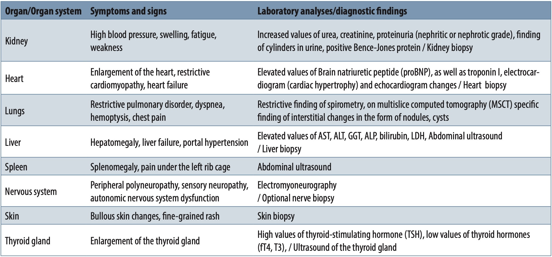
The symptoms and signs of the affected organ dominate the clinical picture of light chain deposition disease. According to literature data, the most frequently affected organ is the kidney in about 85% of patients [14]. In about 35% of patients, involvement of the kidneys is not the only localization, and symptoms and signs of involvement of other organs appear along with kidney disorders. Isolated extrarenal manifestations with preserved renal function are rarely present [4]. Involvement of other branches most often involves involvement of the liver (about 23% of cases), involvement of the heart, lungs, peripheral and central nervous system, skin, gastrointestinal tract, spleen, thyroid gland, and others [12],[15],[16],[17],[18],[19],[20].
DIAGNOSTICS
Diagnosing light chain deposition disease consists of several steps. If light chain disease is suspected based on the clinical picture and manifestations of the disease, the first step is a biopsy of the affected organ, most often the kidneys, but also the liver, heart, lungs, skin, and nerves [21]. Amorphous linear deposits of light chains can be seen on the pathohistological preparation, and light chain amyloidosis is ruled out by negative Congo red staining [7]. Confirmation that it is indeed monoclonal light chains is carried out by determining the concentration of light chains in serum and urine, together with serum and urine protein electrophoresis and immunoelectrophoresis. These tests confirm that the light chains are monoclonal, but whether it is only about the light chains or whether they are synthesized as a paraprotein along with the heavy chains [22] is also examined. The last step in the diagnosis is the examination of the bone marrow by pathohistological analysis, where the number of plasma cells is detected, as well as their characteristics on immunohistochemical staining and fluorescent in situ hybridization (FISH) [4],[21],[23]. Alternatively, immunophenotypic characteristics of plasma cells can be examined by flow cytometry in bone marrow aspirate [2],[4],[21].
COURSE, PROGNOSIS, AND TREATMENT
Light chain deposition disease shows a naturally progressive course with eventual loss of function of the affected organ [24]. In almost 70% of patients suffering from light chain deposition disease, there is an association with plasmacytic dyscrasia (most often multiple myeloma) or some other lymphoproliferative disease [13]. Prognostic parameters that affect the further course of the disease are the age of the patient, the presence of other plasmacytic dyscrasias or lymphoproliferative diseases, as well as the presence of extrarenal disease, which represents a significant negative prognostic parameter [24]. If we consider renal prognostic parameters, the speed of development of renal insufficiency has the most significant importance, while in the case of cardiac involvement, biochemical parameters such as proBNP and troponin I are monitored [25].
The treatment of this entity, if it is associated with multiple myeloma or other plasmacytic lymphoproliferative diseases, is treated according to the protocols for treating these diseases. When it comes to the idiopathic form, there are no official consensuses on therapy [26]. Small groups of treated patients are primarily mentioned in the literature, and controlled studies have not been conducted due to the entity’s rarity. According to available literature data, patients were mostly treated with protocols based on therapies for the treatment of multiple myeloma, supported by high-dose chemotherapy and autologous stem cell transplantation. The assessment of the effect of treatment is based on achieving a hematological response (estimation of monoclonal plasma cells in the bone marrow), measuring the concentration of free light chains in the serum, as well as monitoring the function of the affected organ [14],[26].
The treatment of patients was initially based on the application of corticosteroid therapy, which in the first proven cases was applied as monotherapy, stabilizing the affected organ’s function and achieving a partial hematological therapeutic response [23]. In addition to corticosteroid therapy, combined chemotherapy regimens with vincristine, adriablastin (VAD), or vincristine, melphalan, and cyclophosphamide (VMCP) were used as monotherapy [5],[23]. After that, therapy with thalidomide with or without cyclophosphamide was first applied in addition to corticosteroid therapy in a smaller number of patients. Data on the effectiveness of this therapy are mainly based on individual case reports, where we detect both refractoriness to treatment and the achievement of complete longterm remission [27],[28].
The largest number of patients, according to the literature, have so far been treated with bortezomib-based protocols, namely bortezomib+cyclophosphamide+dexamethasone (VCD) or bortezomib+- dexamethasone (VD), achieving a good hematological response. Complete response (CR) was achieved in about 55.6%, and very good partial response (VGPR) and partial response (PR) in other treated patients [26]. The use of autologous transplantation in treating these patients after the initial treatment leads to a longer and more stable remission, with an increase in complete response to 61.5% of patients [20].
Treatment of light chain deposition disease was also carried out in individual cases using therapeutic modalities with lenalidomide. Literature data speak of many different therapeutic regimens, and lenalidomide was combined with prednisone, dexamethasone, melphalan, and prednisone, as well as dexamethasone and cyclophosphamide. The achieved effects were recorded as PR or VGPR, with response maintenance of 1-4 years [29].
In recent years, data have appeared on patients treated with daratumumab as monotherapy in the initial treatment, in whom long-term hematological remission is achieved and maintained [30]. Daratumumab was also observed in consolidation after treatment with protocols based on bortezomib, where the hematological response was improved in all patients (from PR to VGPR or from VGPR to CR) [31].
Transplantation of the affected organ in case of terminal insufficiency is also mentioned as a supportive therapy in these patients, but in order to carry out this procedure, it is necessary to achieve a hematological response and reduce the concentration of light chains in the serum, because otherwise the function of the transplanted organ will be damaged again [32].
CONCLUSION
Light chain deposition disease is a rare entity and diagnostic challenge in most affected patients. A multidisciplinary approach, including nephrologists, hematologists, pathologists, and subspecialists of other branches in case of extrarenal manifestations, is needed to carry out adequate diagnostics, follow-up, and therapy of patients.
Informations

-
Keywords:
-
Received:
-
Revised:
-
Accepted:
-
Online first:
-
DOI:
-
Cite this article:

Danijela Jovanović
Clinic for Hematology, University Clinical Center Kragujevac
30 Zmaj Jovina Street, 34000 Kragujevac, Serbia
E-mail:
1. Alaggio R, Amador C, Anagnostopoulos I, Attygalle AD, Araujo IBO, Berti E, et al. The 5th edition of the World Health Organization Classification of Haematolymphoid Tumours: Lymphoid Neoplasms. 2022 Jul;36(7):1720-48. doi: 10.1038/s41375-022-01620-2. [CROSSREF]
2. Jimenez-Zepeda VH. Light chain deposition disease: novel biological insights and treatment advances. Int J Lab Hematol. 2012 Aug;34(4):347-55. doi: 10.1111/j.1751-553X.2012.01419.x. [CROSSREF]
3. Randall RE, Williamson WC Jr, Mullinax F, Tung MY, Still WJ. Manifestations of systemic light chain deposition. Am J Med. 1976 Feb;60(2):293-9. doi: 10.1016/0002-9343(76)90440-x. [CROSSREF]
4. Pozzi C, D’Amico M, Fogazzi GB, Curioni S, Ferrario F, Pasquali S, et al. Light chain deposition disease with renal involvement: clinical characteristics and prognostic factors. Am J Kidney Dis. 2003 Dec;42(6):1154-63. doi: 10.1053/j. ajkd.2003.08.040. [CROSSREF]
5. Wang Q, Jiang F, Xu G. The pathogenesis of renal injury and treatment in light chain deposition disease. J Transl Med. 2019 Nov 25;17(1):387. doi: 10.1186/ s12967-019-02147-4. [CROSSREF]
6. Keeling J, Teng J, Herrera GA. AL-amyloidosis and light-chain deposition disease light chains induce divergent phenotypic transformations of human mesangial cells. Lab Invest. 2004 Oct;84(10):1322-38. doi: 10.1038/labinvest.3700161. [CROSSREF]
7. Sicard A, Karras A, Goujon JM, Sirac C, Bender S, Labatut D, et al. Light chain deposition disease without glomerular proteinuria: a diagnostic challenge for the nephrologist. Nephrol Dial Transplant. 2014 Oct;29(10):1894-902. doi: 10.1093/ndt/gfu045. [CROSSREF]
8. Büttner-Herold M, Krieglstein N, Teresa Chuva T, Kaija Minuth K, Frederick Pfister F, Daniel C, et al. Light chain restriction in proximal tubules-implications for light chain proximal tubulopathy. Front Med (Lausanne). 2022 Mar 28;9:723758. doi: 10.3389/fmed.2022.723758. [CROSSREF]
9. Gu X, Herrera GA. Light-chain-mediated acute tubular interstitial nephritis: a poorly recognized pattern of renal disease in patients with plasma cell dyscrasia. Arch Pathol Lab Med. 2006 Feb;130(2):165-9. doi: 10.5858/2006-130-165-LATINA. [CROSSREF]
10. Sethi S, Rajkumar SV, D’Agati VD. The complexity and heterogeneity of monoclonal immunoglobulin-associated renal diseases. J Am Soc Nephrol. 2018 Jul;29(7):1810-23. doi: 10.1681/ASN.2017121319. [CROSSREF]
11. Mohan M, Buros A, Mathur P, Gokden N, Singh M, Susanibar S, et al. Clinical characteristics and prognostic factors in multiple myeloma patients with light chain deposition disease Am J Hematol. 2017 Aug;92(8):739-45. doi: 10.1002/ajh.24756. [CROSSREF]
12. Gandhi M, Pasha SB, Reznicek E, Pasha SR, Ertugrul H, Araslanova A, et al. A case of light chain deposition disease leading to acute liver failure and review of literature. Diseases. 2023 Feb 1;11(1):24. doi: 10.3390/diseases11010024. [CROSSREF]
13. Kanzaki G, Okabayashi Y, Nagahama K, Ohashi R, Tsuboi N, Yokoo T, et al. Monoclonal immunoglobulin deposition disease and related diseases. J Nippon Med Sch. 2019;86(1):2-9. doi: 10.1272/jnms.JNMS.2019_86-1. [CROSSREF]
14. Cassano Cassano R, Bonadio AG, Del Giudice ML, Giannese D, Galimberti S, Buda G. Light chain deposition disease: pathogenesis, clinical characteristics and treatment strategies. Ann Hematol. 2024 Aug 28. doi: 10.1007/s00277- 024-05911-9. [CROSSREF]
15. Colombat M, Gounant V, Mal H, Callard P, Milleron B. Light chain deposition disease involving the airways: diagnosis by fibreoptic bronchoscopy. Eur Respir J. 2007 May;29(5):1057-60. doi: 10.1183/09031936.00134406. [CROSSREF]
16. Hudak M, Sardana R, Parwani AV, Mathewson RC, Gibson CG, Cohen PA, et al. Light chain deposition disease presenting as an atrial mass: a case report and review of literature. Cardiovasc Pathol. 2021 Nov-Dec;55:107368. doi: 10.1016/j.carpath.2021.107368. [CROSSREF]
17. Grassi MP, Clerici F, Perin C, Borella M, Gendarini A, Quattrini A, et al. Light chain deposition disease neuropathy resembling amyloid neuropathy in a multiple myeloma patient. Ital J Neurol Sci. 1998 Aug;19(4):229-33. doi: 10.1007/BF02427609. [CROSSREF]
18. Dos Santos LP, Couto J, Romano M, López R. Light-chain deposition disease presenting with spontaneous splenic rupture. Eur J Case Rep Intern Med. 2018 Dec 27;5(12):0001010. doi: 10.12890/2018_0001010. [CROSSREF]
19. Hendricks C, Fernández Figueras MT, Liersch J, Martin-Urdà MT, López D, Brochhausen C, et al. cutaneous light chain deposition disease: a report of 2 cases and review of the literature. Am J Dermatopathol. 2018 May;40(5):337- 41. doi: 10.1097/DAD.0000000000000991. [CROSSREF]
20. Hiyamuta H, Yamada S, Matsukuma Y, Tsuchimoto A, Nakano T, Taniguchi M, et al. Light chain deposition disease in an older adult patient successfully treated with long-term administration of bortezomib, melphalan and prednisone. Intern Med. 2016;55(10):1319-25. doi: 10.2169/internalmedicine.55.5752. [CROSSREF]
21. Gavriatopoulou M, Musto P, Caers J, Merlini G, Kastritis E, van de Donk N, et al. European myeloma network recommendations on diagnosis and management of patients with rare plasma cell dyscrasias. Leukemia. 2018 Sep;32(9):1883-98. doi: 10.1038/s41375-018-0209-7. [CROSSREF]
22. Yadav P, Leung N, Sanders PW, Cockwell P. The use of immunoglobulin light chain assays in the diagnosis of paraprotein-related kidney disease. Kidney Int. 2015 Apr;87(4):692-7. doi: 10.1038/ki.2014.333. [CROSSREF]
23. Gertz MA. Immunoglobulin light chain amyloidosis: 2018 Update on diagnosis, prognosis, and treatment. Am J Hematol. 2018 Sep;93(9):1169-80. doi: 10.1002/ajh.25149. [CROSSREF]
24. Sayed RH, Wechalekar AD, Gilbertson JA, Bass P, Mahmood S, Sachchithanantham S, et al. Natural history and outcome of light chain deposition disease. Blood. 2015 Dec 24;126(26):2805-10. doi: 10.1182/blood-2015-07-658872. [CROSSREF]
25. Li XM, Rui HC, Liang DD, Xu F, Liang SS, Zhu XD, et al. Clinicopathological characteristics and outcomes of light chain deposition disease: an analysis of 48 patients in a single Chinese center. Ann Hematol. 2016 May;95(6):901-9. doi: 10.1007/s00277-016-2659-1. [CROSSREF]
26. Masood A, Ehsan H, Iqbal Q, Salman A, Hashmi H. Treatment of light chain deposition disease: a systematic review. J Hematol. 2022 Aug;11(4):123-30. doi: 10.14740/jh1038. [CROSSREF]
27. Wang D, Wang Y, Sun S. Renal pathological changes after successful treatment of LCDD using cyclophosphamide, thalidomide, and dexamethasone. Ren Fail. 2021 Dec;43(1):1425-7. doi: 10.1080/0886022X.2021.1988967. [CROSSREF]
28. Fujita H, Hishizawa M, Sakamoto S, Kondo T, Kadowaki N, Ishikawa T, et al. Durable hematological response and improvement of nephrotic syndrome on thalidomide therapy in a patient with refractory light chain deposition disease. Int J Hematol. 2011 May;93(5):673-6. doi: 10.1007/s12185-011- 0829-4. [CROSSREF]
29. Kimura S, Ohkawara H, Ogawa K, Tanaka M, Sano T, Harada-Shirado K, et al. Lenalidomide as a beneficial treatment option for renal impairment caused by light chain deposition disease. Intern Med. 2018 Dec 15;57(24):3651-7. doi: 10.2169/internalmedicine.1018-18. [CROSSREF]
30. Milani P, Basset M, Curci P, Foli A, Rizzi R, Nuvulone M, et al. Daratumumab in light chain deposition disease; rapid and profound hematologic response preserves kidney function. Blood Adv. 2020 Apr14;4(7):1321-4. doi: 10.1182/ bloodadvances.2020001553. [CROSSREF]
31. Kastritis E, Rousakis P, Kostopoulos IV, Gavriatopoulou M, Theodorakakou F, Fotiou D, et al. Consolidation with a short course of daratumumab in patients with AL amyloidosis or light chain deposition disease. Amyloid. 2021 Dec;28(4):259-66. doi: 10.1080/13506129.2021.1971192. [CROSSREF]
32. Heybeli C, Alexander MP, Bentall AJ, Amer H, Buadi FK, Dean PG, et al. Kidney transplantation in patients with monoclonal gammopathy of renal significance (MGRS)-associated lesions: a case series. Am J Kidney Dis. 2022 Feb;79(2):202-16. doi: 10.1053/j.ajkd.2021.04.015. [CROSSREF]