


Postoperative complications and their correlation with the surgical technique in the treatment of gastroschisis
- University Clinical Center of Serbia, Clinic for Hematology, Belgrade, Serbia
- Mother and Child Health Institute of Serbia Dr Vukan Čupić, Belgrade, Serbia
ABSTRACT
Introduction: Gastroschisis is a congenital paraumbilical defect of the anterior abdominal wall with evisceration of the abdominal organs. A modern approach to treating gastroschisis has contributed to better results, as evidenced by the 5% – 10% mortality rate.
Aim: The study aims to evaluate the frequency of complications and death in the population due to gastroschisis, as well as the connection between the surgical tech- niques used in the treatment and the outcome of the treatment.
Material and methods: The retrospective cohort study included 75 patients diag- nosed with gastroschisis, treated from 2000 to 2020 at the Mother and Child Health Institute of Serbia Dr Vukan Čupić. By applying the exclusion criteria, a sample of 61 patients was obtained. Two cohorts of subjects were formed based on the applied surgical method, namely: primary repositioning and fascial closure of the defect (gastroschisis surgical cohort – GSC), i.e. delayed primary repositioning of the defect using a silastic bag (silastic gastroschisis cohort – SGC).
Results: The gastroschisis surgical cohort comprised 38 patients, while the silastic gas- troschisis cohort comprised 23 subjects. It was found that necrotizing enterocolitis (NEC) was a statistically significantly more frequent complication in subjects from the silastic gastroschisis cohort (5/23, i.e. 21.7% in SGC and 0/38 in GSC; RR 0.32, 95% CI: 0.22 – 0.47; p = 0.003). The statistical significance of the difference in relation to the frequency of other complications was not proven: ileus (0/23 in SGC and 5/38, i.e. 13.2% in GSC; RR 0.59, 95% CI: 0.47 - 0.73, p = 0.069), compartment syndrome (0/23 in SGC and 2/38, i.e. 5.3% in GSC; RR 0.61, 95% CI: 0.50 – 0.75, p = 0.263) and death ( 2/23, i.e. 8.7% in SGC and 2/38, i.e. 5.3% in GSC; RR 1.26, 95% CI: 0.46 – 3.43, p = 0.600).
Conclusion: There is no distinctive proof of the superiority of one method over an- other. The risks of ileus and compartment syndrome are higher when applying pri- mary fascial closure of the defect, while the risks of NEC and fatal outcome are higher when the silo method (use of a silastic bag, i.e., silo bag) is applied. The choice of method in treating gastroschisis depends on the abdominovisceral disproportion and the physical appearance of the eviscerated intestines, assessed with the Gastroschisis Prognostic Score (GPS).
INTRODUCTION
Gastroschisis is a congenital paraumbilical defect of the anterior abdominal wall with evisceration of abdominal organs. It occurs in 1 – 4 : 10,000 newborns [1]. It happens more often in males and premature infants. Unlike other anterior abdominal wall defects, gastroschisis is not usually associated with chromosomal or other structural anomalies, except for intestinal atresia or stenosis, which are seen in about 10% of cases. Usually, small and large intestine loops are eviscerated, however, a part of the stomach, ovaries, fallopian tubes, testicles, and urinary bladder can also be involved. Because of the evisceration of the organs, there is an increased loss of fluid and a susceptibility of the newborn to hypothermia. The intestines are glossy in appearance, with thickened walls. They are shortened, covered in exudate, with adhesions between the loops. Signs of irritative peritonitis occur due to the exposure of the intestine to the amniotic fluid. One of the primary problems of gastroschisis is the relatively underdeveloped abdominal cavity, which cannot accommodate the eviscerated organs without a significant increase in intra-abdominal pressure. The propensity of the paraumbilical defect to contract significantly contributes to abnormal vascularization of the eviscerated intestines [2],[3].
The contemporary approach to the surgical treatment of gastroschisis has contributed to better results, which is evident from the mortality rate that does not exceed 5% – 10% [4]. Complicated gastroschisis (associated with atresia, volvulus, ischemia, stenosis, necrosis, and intestinal perforation) has a poor outcome, with a mortality rate of 19% – 30%. The traditional approach to the treatment of gastroschisis involves primary manual repositioning of the eviscerated organs and double-layered suturing of the defect (fascia and skin), under general anesthesia. Delayed primary or staged repositioning is necessary when the surgeon cannot reposition organs without a resulting significant increase in intra-abdominal pressure. In such cases, a silo bag with a reinforced loop is applied, enabling gravitational repositioning of the intestines [5],[6],[7].
The most significant life-threatening complication of primary manual repositioning of eviscerated organs and fascial suturing of the defect is the development of abdominal compartment syndrome, which is defined as a prolonged increase in intraperitoneal pressure above 20 mmHg (the normal pressure is around 6.5 mmHg), with the development of multiorgan dysfunction [8],[9],[10]. Risk factors include the following: reduced compliance of the abdominal wall, increased intestinal intraluminal volume, increased abdominal volume, and increased capillary permeability [11]. Abdominal compartment syndrome is defined by the presence of the following clinical signs: increased systemic and pulmonary vascular resistance, oliguria, hypoxia, decreased cardiac output, hypotension, and acidosis [12].
Abdominal hypertension is the cause of the development of ischemia, massive necrosis, intestinal perforation, renal and respiratory insufficiency, and sepsis, and by extension, it results in the cause-and-effect relationship in the development of systemic inflammatory response syndrome (SIRS) and multiple organ dysfunction syndrome (MODS) [8],[13],[14]. In addition, elevated intra-abdominal pressure can lead to gastroesophageal reflux, intolerance of enteral intake, malabsorption, and pseudo-obstruction. Intolerance and malabsorption are common due to the pathophysiological nature of the relatively short intestine, while the development of pseudo-obstruction, which can further progress to ileus, manifests as distension of the abdomen, dilation of the intestinal loops with intestinal stasis, an increase in the number of bacteria, and the development of infection, and even sepsis [15].
Another significant factor of morbidity and mortality in the population with gastroschisis is the development of necrotizing enterocolitis (NEC). Literature shows that typical NEC, including NEC associated with gastroschisis, is, in fact, the direct consequence of the typical characteristics of the patient, i.e., preterm infants of low gestational age and low birth weight [16] with signs of asphyxia and hypovolemia, exhibiting transmural ischemic changes in the intestines and consequently direct translocation of bacteria into the lymph flow and blood circulation, which can lead to increased SIRS, as well as to MODS [17],[18]. Recent literature data, pertaining to the last five years, indicate that the overall incidence of NEC has decreased, as compared to earlier reports, i.e., from 15% – 20% [19] to 3% – 5%, as it is today [16],[20],[21]. NEC in gastroschisis does not have its typical pathophysiological mechanisms, it differs from typical NEC because it is delayed, occurs later, and is more often a consequence of postoperative compartment syndrome, i.e., of compromised intestinal circulation and ischemia, but also of delayed postoperative initiation of enteral nutrition, or of prolonged starvation associated with the excessive multiplication of bacteria and the development of an infection, which is especially dangerous if it is of clostridial origin. Timely enteral intake of breast milk is believed to be protective and to reduce the risk of NEC after operative treatment of gastroschisis [21],[22],[23].
On the other hand, exteriorized intestines, with associated malrotation and mesenterium commune, are prone to torsion and volvulus of the midgut, which can lead to mesenteric circulation disorders, decreased blood flow, and gangrene of the affected intestine. Sometimes, the excessive tightness of the blood vessels of the mesenterium, compression by the edges of the defect on the front abdominal wall, or a tight silo bag can be enough to compromise the mesenteric circulation. Intestinal loops may require unwinding and appropriate placing in the abdomen or silo bag to avoid strangulation and gangrene, and if the abdominal defect is small, as compared to the mass of the exteriorized intestines, it is necessary to dilate it immediately, to avoid vascular compromise [24].
The treatment of complex forms of gastroschisis is accompanied by numerous complications: the need for repeated laparotomies, ultra-short bowel syndrome, long-term parenteral nutrition, cholestatic liver failure, sepsis, and high treatment costs [5],[25].
The study aims to assess the frequency of complications and fatal outcome in the population with gastroschisis and to assess the association between the surgical techniques applied in the treatment of gastroschisis and the occurrence of the said complications, i.e., fatal outcome.
MATERIALS AND METHODS
The studied population of patients with gastroschisis consisted of 75 patients. Based on the exclusion criteria (ischemia, stenosis, necrosis, perforation of the intestine present at birth, presence of intestinal atresia, agenesis of the small intestine and colon, a small defect of the anterior abdominal wall wherein the silo bag cannot be placed), a selection was made, wherefore the study did not apply to 14 (14/75) respondents. Thereby, 61 (61/75) respondents remained in the study sample.
This is a retrospective cohort study. The study sample consists of newborns diagnosed with gastroschisis, treated between 2000 and 2020, at the Mother and Child Health Institute of Serbia Dr Vukan Čupić. This is the reference hospital for prenatal diagnosis and treatment of this congenital anomaly.
Data for analysis were collected from medical records, existing permanent medical histories that contain all the data collected from patient admission to the Department of Surgical Neonatal Intensive Care until the time of discharge or transfer to the Department of Pediatric Intensive Care.
Until December 2006, the surgical technique of manual repositioning of eviscerated organs and primary fascial closure of the defect under general anesthesia was applied. The group of patients treated in this way consisted of 38 patients comprising the gastroschisis surgical cohort (GSC). As of January 1, 2007, a new approach has been applied – delayed primary closure of the defect without general anesthesia, i.e. gravitational repositioning using a silo bag with a reinforced loop. This group consisted of 23 patients comprising the silastic gastroschisis cohort (SGC). Variables related to the characteristics of gastroschisis and the choice of surgical technique were determined by the type of complication: compartment syndrome, ileus, NEC, and fatal outcome.
The data collected were entered into a computer database, and the SPSS 2016 program was used for data processing. The assessment of the frequency of complications and fatal outcome was given in the form of a 95% confidence interval (CI). The significance of the difference was tested with the parametric Student t-test (for normal data distribution) and the Mann-Whitney U test (for non-normal data distribution). Data were considered statistically significant for p < 0.05. The attributable risk (AR) and the the relative risk (RR) were used to assess the association between surgical techniques and complications, as an outcome. The results are presented textually, tabularly, and graphically.
RESULTS
Data related to the frequency and rate of complications are presented in Table 1. NEC was registered in 5 out of 23 (21.7%) patients, in whom the silo bag closure technique was applied, while no cases of NEC were registered with surgical closure (0/ 38). Ileus developed in 5 out of 38 (13.2%) patients treated with the surgical technique, while this complication did not occur in the second cohort (0/23 in SGC). Compartment syndrome developed in two out of 38 (5.3%) patients treated with primary fascial closure, but no cases of this complication were registered in the cohort where closure of the defect with a silo bag was applied (0/23 in SGC). Fatal outcome was registered in two out of 38 (5.3%) patients in the gastroschisis surgical cohort and two out of 23 (8.7%) patients in the silastic gastroschisis cohort.
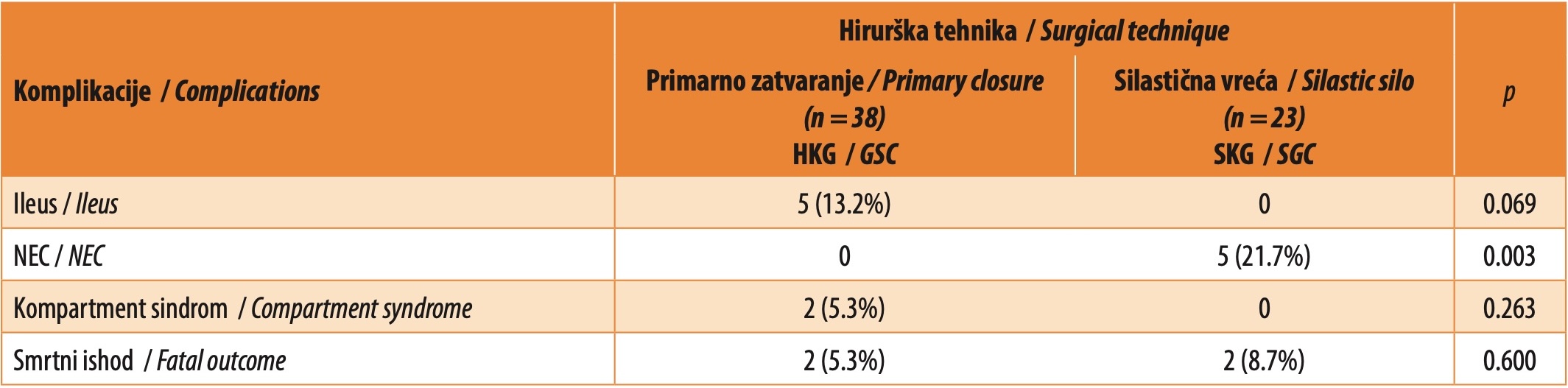
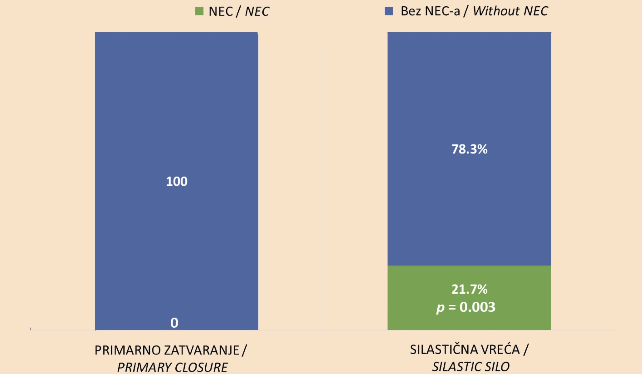
The conclusion is that there was no statistically significant difference between primary repositioning and fascial closure of the defect, in relation to the application of a silo bag, and in terms of the frequency of the occurrence of ileus (p = 0.069), compartment syndrome (p = 0.263) and fatal outcome (p = 0.600). On the other hand, it was inferred that there was a statistically significant difference between primary repositioning and fascial closure of the defect and the application of a silo bag, as to the frequency of NEC. NEC was a statistically significantly more frequent complication in subjects of the silastic gastroschisis cohort (p = 0.003). This statistical significance is shown in Figure 1.
To assess the association between surgical techniques and complications and fatal outcome, the attributable (absolute) risk (AR) and the relative risk (RR) were used, presented as a 95% confidence interval (95% CI). These data are presented in Table 2. It was concluded that the attributable risk of ileus resulting from exposure to a certain surgical technique, was 13% in GSC and 0% in SGC, with a 95% CI. The attributable risk of ileus was 13% in the study sample and was associated with the primary fascial closure technique, with a 95% CI. In the study sample, the relative risk of developing ileus was 0.59 times higher in the surgical cohort than in the silastic gastroschisis cohort, with a 95% CI. The relative risk of ileus in the population was 0.47 – 0.73 times higher when the technique of primary fascial closure of the defect was applied, with a 95% CI.
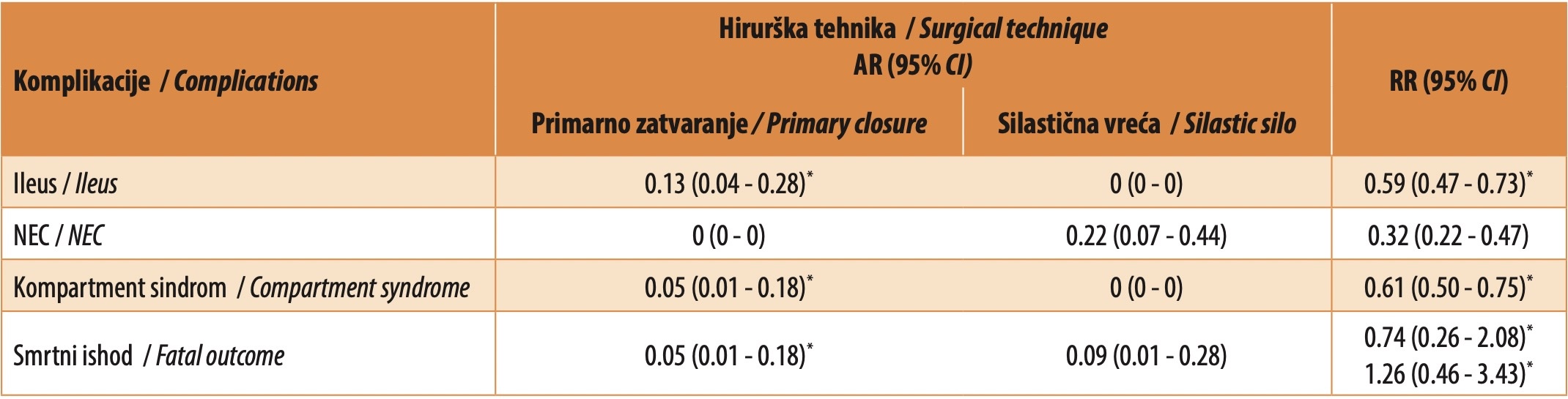
The attributable risk of NEC due to exposure to a particular surgical technique was 0% in GSC and 22% in SGC, with a 95% CI. The attributable risk of NEC in the study sample was 22% and was associated with the silastic bag application technique, with a 95% CI. In the population, the incidence of NEC, as a complication in patients with gastroschisis, was 4% – 18.5% [22]. The relative risk of developing NEC was 0.32 times higher in SGC than in GSC, with a 95% CI. In the population, the relative risk of developing NEC was 0.22 – 0.47 times higher with the application of the silo bag technique than with the application of primary manual repositioning and fascial closure of the defect, with a 95% CI.
The attributable risk of compartment syndrome caused by exposure to a particular surgical technique was 5% in GSC and 0% in SGC, with a 95% CI. The attributable risk of compartment syndrome in the study sample was 5%, and was the consequence of the application of primary repositioning of eviscerated organs and fascial closure of the defect, with a 95% CI. The relative risk of the occurrence of compartment syndrome was 0.61 times higher in GSC, as compared to SGC, with a 95% CI. In the population, the relative risk of compartment syndrome was 0.50 – 0.75 times higher when applying the technique of primary repositioning of eviscerated organs and fascial closure of the defect than when applying the silo bag technique, with a 95% CI.
The attributable risk of fatal outcome resulting from exposure to a particular surgical technique was 5% in GSC, and 9% in SGC, with a 95% CI. In the population, the absolute risk of fatal outcome due to complications, which was associated with the technique of primary repositioning of eviscerated organs and fascial closure of the defect, was 1% – 18%, with a 95% CI, while the absolute risk associated with the silo bag application technique was 1% – 28%, with a 95% CI. The relative risk of fatal outcome was 1.26 times higher in SGC than in GSC, with a 95% CI. In the population, the relative risk of death as a complication was 0.46 – 3.43 times higher in SGC than in GSC, with a 95% CI. However, in the study sample, there was no statistical significance in the difference between the silastic gastroschisis cohort and the gastroschisis surgical cohort, as to the frequency of fatal outcome.
DISCUSSION
Appropriate treatment of gastroschisis requires the integration of several disciplines (surgery, gynecology and obstetrics, neonatology, pediatrics). Gastroschisis can be diagnosed at regular follow-up ultrasound examinations during pregnancy, as well as by determining the mother’s serum alpha-fetoprotein level (screening), which indicates the utmost importance of prenatal care. In developed countries, the number of gastroschisis cases detected in this way exceeds 90%. There are two important questions arising in perinatology: how and when is the best way to end this kind of pregnancy? These questions are the subject of many studies.
In their study, O’Connell et al. [26] recommend delivery at 37 weeks of pregnancy, noting that a cesarean section remains reserved only for pregnant women who have the usual indications for it. Once the baby is born and placed in the intensive care unit, the initial therapeutic goals are to maintain homeostasis through intravenous fluid replacement, respiratory support, thermoregulation, and bowel protection [27]. This study deals with the analysis of the surgical treatment of gastroschisis and the association between the applied surgical techniques and the occurrence of complications.
The optimal surgical approach is still the subject of many studies. Regardless of the technique used, the goal of the surgical treatment of gastroschisis is to reposition the intestine and close the abdomen while avoiding an increase in intra-abdominal pressure above physiological levels. This can be achieved immediately after birth by primary manual bowel repositioning and suturing of the fascial defect or with delayed bowel repositioning achieved by the placement of a silo bag and subsequent closure of the defect. Primary closure is not always possible and safe, especially when abdominovisceral disproportion exists, which is when there is a possibility of abdominal compartment syndrome. This potentially fatal complication requires immediate opening of the abdomen. Thus, the silo technique was initially used only in cases that were not amenable to primary closure. However, many sources advocate the use of both techniques. Numerous studies indicate that their success rate is generally equal [28]. Recent research emphasizes the importance of the time elapsed from birth to surgery – optimally it is less than three hours [29].
In our study sample, the technique of primary repositioning and fascial closure of the defect was applied in 62.3% (38/61) of the subjects, while a silo bag with a reinforced loop was used in 37.7% (23/61) of the patients. The incidence of NEC was statistically significantly more frequent in SGC. The frequency rate of NEC was 21.7% (5/23) in subjects belonging to the SGC, while in subjects belonging to the GSC, no NEC was recorded. The absolute risk of NEC in the study sample was 22% and it was associated with the silo bag application technique (in the population, the AR ranged from 7% – 44%). The risk of developing NEC was 0.32 times greater in SGC than in GSC, with a 95% confidence interval (RR 0.32, 95% CI: 0.22 – 0.47, p = 0.003).
Our study shows that delayed primary closure of the defect is associated with a higher risk of NEC, although the pathophysiology of NEC associated with the silastic bag technique is not entirely clear. Although complications of intestinal ischemia and gangrene could be expected when applying this surgical method in the treatment of gastroschisis, the reason may be that it is applied more often, due to the small abdominal cavity, in premature children, who are of small body weight and prone to developing NEC.
Also, the results of our study suggest that patients treated with primary repositioning of eviscerated organs and fascial closure of the defect may have better outcomes because they have avoided the risk of NEC. Furthermore, avoiding elective preterm deliveries could reduce the incidence of NEC in patients with gastroschisis [30].
For other complications, the statistical significance of the difference was not proven – ileus (RR 0.59, 95% CI: 0.47 - 0.73, p = 0.069) and compartment syndrome (RR 0.61, 95% CI: 0.50 - 0 .75, p = 0.263), although the risk of their occurrence was higher in the gastroschisis surgical cohort when applying the method of primary repositioning of eviscerated organs and fascial closure of the defect, while the risk of fatal outcome (RR 1.26, 95% CI: 0.46 - 3.43, p = 0.600) was higher in the silastic gastroschisis cohort, when applying the silo technique in delayed primary care of gastroschisis.
Despite the existing results, it is difficult to say which method will give a better outcome. While one meta-analysis indicates that primary fascial closure gives better results [16], another shows that patients in whom a silo bag is used remain on mechanical ventilation for a shorter period, but require longer total parenteral nutrition [31].
In a 2020 paper by Raymond et al. [32], it was reported that the overall survival of patients with gastroschisis until discharge from hospital was an incredible 95%. However, the frequency of reoperations after the neonatal period is significant, because approximately 25% of these patients undergo surgical treatment during the first year of life, due to intestinal obstruction.
Wissanji et al. [33] explain that the Gastroschisis Prognostic Score (GPS) is currently the best available tool for a quick and reliable assessment of the severity of the disease. This system was created in 2012 by Cowan et al. It can be used to determine the degree of exposure of eviscerated organs to amniotic fluid, as well as to evaluate the method of choice and the outcome of treatment, based only on the appearance of the intestine. GPS scoring and calculation must be done immediately after birth, using a standardized visual scoring system, which is determined by four elements: ‘glossy’ intestines, the presence of necrosis, atresia, and/or perforation. Patients can be classified into two categories: low-risk patients (GPS ≤ 1) and high-risk patients (GPS ≥ 2) [34],[35].
If the assessment of the appearance of the intestines shows the presence of a ‘peel’, i.e., ‘rind’ on the intestines, and there is also an abdominovisceral disproportion, this is an indication for the placement of a silastic bag [36]. Although the indications are clear, the placement of the silastic bag should be approached with caution, because if the opening on the anterior abdominal wall is too small (< 1.5 cm), the vascularization may be further compromised and lead to intestinal ischemia and further complications. Therefore, it is up to the surgeon to assess whether it is necessary to perform an additional fasciotomy before placing the silo bag so that the opening on the anterior abdominal wall is of the appropriate size. It is precisely in this assessment that an error may occur, which is why there is a more frequent association of NEC and fatal outcome with the use of a silo bag in the treatment of gastroschisis.
In our study, the answers to many questions remain unanswered. The reason for this is the small sample of respondents, which is why the research itself does not have great statistical power. The size of the sample is consistent with the fact that gastroschisis is a rare congenital anomaly.
CONCLUSION
There is no distinctive proof of the superiority of one method in the treatment of gastroschisis over another. The study found that the risks of ileus and compartment syndrome are higher when the technique of primary fascial closure of the defect is applied, as well as that the risk of NEC and fatal outcome is higher when using the silo method. The choice of the treatment method depends on the presence or absence of abdominovisceral disproportion and the physical appearance of the eviscerated intestines, assessed with GPS. Based on the studies published so far, it can be said that success in the treatment of gastroschisis is high and that special attention needs to be paid to perioperative treatment and surgical technique. However, future multicentric studies are necessary for a systematic review and statistically significant results.
Informations

-
Keywords:
-
Received:
-
Revised:
-
Accepted:
-
Online first:
-
DOI:
-
Cite this article:

Natalija Kecman
Clinic for Hematology, University Clinical Center of Serbia
2 Dr Koste Todorovića Street, 11000 Belgrade, Serbia
E-mail:
1. Klein MD. Congenital defects of the abdominal wall. In: Grosfeld JL, O’Neill JA, Fonkalsrud EW, Coran AG, editors. Pediatric Surgery. 6th ed. Philadelphia: Mosby Elsevier, 2006. p. 1157-1171. [CROSSREF]
2. Bruch SW, Langer JC. Omphalocele and gastroschisis. In: Pre Puri, editor. Newborn surgery. 3rd ed. London: Arnold, 2010. p. 605-611.
3. Moore TC, Stokes GE. Gastroschisis. Surgery 1953; 33:112-115.
4. Safavi A, Skarsgard ED. Advances in the surgical treatment of gastroschisis. Surg Technol 2015; 2637-41.
5. Arnold MA, Chang DC, Nabaweesi R, Colombani PM, Bathurst MA, Mon KS, et al. Risk stratification of 4344 patients with gastroschisis into simple and complex categories. J Pediatr Surg. 2007 Sep;42(9):1520-5. doi: 10.1016/j. jpedsurg.2007.04.032. [CROSSREF]
6. Abdullah F, Arnold MA, Nabaweesi R, Fischer AC, Colombani PM, Anderson KD, et al. Gastroschisis in the United States 1988-2003: analysis and risk categorization of 4344 patients. J Perinatol. 2007 Jan;27(1):50-5. doi: 10.1038/ sj.jp.7211616. [CROSSREF]
7. Lobo JD, Kim AC, Davis RP, Segura BJ, Alpert H, Teitelbaum DH, et al. No free ride? The hidden costs of delayed operative management using a spring-loaded silo for gastroschisis. J Pediatr Surg. 2010 Jul;45(7):1426-32. doi: 10.1016/j.jpedsurg.2010.02.047. [CROSSREF]
8. David AL, Tan A, Curry J. Gastroschisis: sonographic diagnosis, associations, management and outcome. Prenat Diagn. 2008 Jul;28(7):633-44. doi: 10.1002/pd.1999. [CROSSREF]
9. De Waele JJ. Abdominal Compartment Syndrome in Severe Acute Pancreatitis - When to Decompress? Eur J Trauma Emerg Surg. 2008 Feb;34(1):11-6. doi: 10.1007/s00068-008-7170-5. [CROSSREF]
10. Burch JM, Moore EE, Moore FA, Franciose R. The abdominal compartment syndrome. Surg Clin North Am. 1996 Aug;76(4):833-42. doi: 10.1016/s0039- 6109(05)70483-7. [CROSSREF]
11. Cheatham ML, Malbrain ML, Kirkpatrick A, Sugrue M, Parr M, De Waele J, et al. Results from the International Conference of Experts on Intra-abdominal Hypertension and Abdominal Compartment Syndrome. II. Recommendations. Intensive Care Med. 2007 Jun;33(6):951-62. doi: 10.1007/s00134-007-0592-4. [CROSSREF]
12. Tal R, Lask DM, Keslin J, Livne PM. Abdominal compartment syndrome: urological aspects. BJU Int. 2004 Mar;93(4):474-7. doi: 10.1111/j.1464- 410x.2003.04654.x. [CROSSREF]
13. Bailey J, Shapiro MJ. Abdominal compartment syndrome. Crit Care. 2000;4(1):23-9. doi: 10.1186/cc646. [CROSSREF]
14. Walker J, Criddle LM. Pathophysiology and management of abdominal compartment syndrome. Am J Crit Care. 2003 Jul;12(4):367-71. [CROSSREF]
15. Asz-Sigall J, Ramirez-Resendiz A, Assia-Zamora S, Lopez-Zertuche-Ortiz JP, Medina-Vega FA. Necrotizing enterocolitis manifesting with pneumatosis ani in a patient with gastroschisis. J Ped Surg Case Reports 2015; 237-238. doi: 10.1016/j.epsc.2015.03.015. [CROSSREF]
16. Schlueter R, Abdessalam S, Raynor S, Cusick R. Necrotizing Enterocolitis following Gastroschisis Repair: An Update. Graduate Medical Education Graduate Medical Education Research Journal Research Journal [Internet]. 2019;1(1). Dostupno na: https://digitalcommons.unmc.edu/gmerj/vol1/iss1/5 [HTTP]
17. Bennet L, Quaedackers JS, Gunn AJ, Rossenrode S, Heineman E. The effect of asphyxia on superior mesenteric artery blood flow in the premature sheep fetus. J Pediatr Surg. 2000 Jan;35(1):34-40. doi: 10.1016/s0022- 3468(00)80009-3. [CROSSREF]
18. Cullen DJ, Coyle JP, Teplick R, Long MC. Cardiovascular, pulmonary, and renal effects of massively increased intra-abdominal pressure in critically ill patients. Crit Care Med. 1989 Feb;17(2):118-21. doi: 10.1097/00003246- 198902000-00002. [CROSSREF]
19. Jayanthi S, Seymour P, Puntis JW, Stringer MD. Necrotizing enterocolitis after gastroschisis repair: a preventable complication? J Pediatr Surg. 1998 May;33(5):705-7. doi: 10.1016/s0022-3468(98)90191-9. [CROSSREF]
20. Fullerton BS, Velazco CS, Sparks EA, Morrow KA, Edwards EM, Soll RF, et al. Contemporary Outcomes of Infants with Gastroschisis in North America: A Multicenter Cohort Study. J Pediatr. 2017 Sep;188:192-197.e6. doi: 10.1016/j. jpeds.2017.06.013. [CROSSREF]
21. Tannuri AC, Sbragia L, Tannuri U, Silva LM, Leal AJ, Schmidt AF, et al. Evolution of critically ill patients with gastroschisis from three tertiary centers. Clinics (Sao Paulo). 2011;66(1):17-20. doi: 10.1590/s1807-59322011000100004. [CROSSREF]
22. Schwartz MZ, Timmapuri SJ. Gastroschisis. In: Puri, P. (eds) Pediatric Surgery. Springer, Berlin, Heidelberg, 2020;1177-87. https://doi.org/10.1007/978-3-662-43588-5_84. [CROSSREF]
23. Riggle KM, Davis JL, Drugas GT, Riehle KJ. Fatal Clostridial necrotizing enterocolitis in a term infant with gastroschisis. J Ped Surg Case Reports 2016; 14:29-31. https://doi.org/10.1016/j.epsc.2016.08.007. [CROSSREF]
24. Gharpure V. Gastroschisis. J Neonatal Surg. 2012 Oct 1;1(4):60. [CROSSREF]
25. Kumar T, Vaughan R, Polak M. A proposed classification for the spectrum of vanishing gastroschisis. Eur J Pediatr Surg. 2013 Feb;23(1):72-5. doi: 10.1055/ s-0032-1330841. [CROSSREF]
26. O’Connell RV, Dotters-Katz SK, Kuller JA, Strauss RA. Gastroschisis: A Review of Management and Outcomes. Obstet Gynecol Surv. 2016 Sep;71(9):537-44. doi: 10.1097/OGX.0000000000000344. [CROSSREF]
27. Haddock C, Skarsgard ED. Understanding gastroschisis and its clinical management: where are we? Expert Rev Gastroenterol Hepatol. 2018 Apr;12(4):405-415. doi: 10.1080/17474124.2018.1438890. [CROSSREF]
28. Skarsgard ED. Management of gastroschisis. Curr Opin Pediatr. 2016 Jun;28(3):363-9. doi: 10.1097/MOP.0000000000000336. [CROSSREF]
29. Allin BS, Tse WH, Marven S, Johnson PR, Knight M. Challenges of improving the evidence base in smaller surgical specialties, as highlighted by a systematic review of gastroschisis management. PLoS One. 2015 Jan 26;10(1):e0116908. doi: 10.1371/journal.pone.0116908. [CROSSREF]
30. Jaczyńska R, Mydlak D, Mikulska B, Nimer A, Maciejewski T, Sawicka E. Perinatal Outcomes of Neonates with Complex and Simple Gastroschisis after Planned Preterm Delivery-A Single-Centre Retrospective Cohort Study. Diagnostics (Basel). 2023 Jun 30;13(13):2225. doi: 10.3390/diagnostics13132225. [CROSSREF]
31. Ross AR, Eaton S, Zani A, Ade-Ajayi N, Pierro A, Hall NJ. The role of preformed silos in the management of infants with gastroschisis: a systematic review and meta-analysis. Pediatr Surg Int. 2015 May;31(5):473-83. doi: 10.1007/ s00383-015-3691-2. [CROSSREF]
32. Raymond SL, Hawkins RB, St Peter SD, Downard CD, Qureshi FG, Renaud E, et al. Predicting Morbidity and Mortality in Neonates Born With Gastroschisis. J Surg Res. 2020 Jan;245:217-224. doi: 10.1016/j.jss.2019.07.065. [CROSSREF]
33. Wissanji H, Puligandla PS. Risk stratification and outcome determinants in gastroschisis. Semin Pediatr Surg. 2018 Oct;27(5):300-303. doi: 10.1053/j. sempedsurg.2018.08.007. [CROSSREF]
34. Cowan KN, Puligandla PS, Laberge JM, Skarsgard ED, Bouchard S, Yanchar N, et al.; Canadian Pediatric Surgery Network. The gastroschisis prognostic score: reliable outcome prediction in gastroschisis. J Pediatr Surg. 2012 Jun;47(6):1111-7. doi: 10.1016/j.jpedsurg.2012.03.010. [CROSSREF]
35. Puligandla PS, Baird R, Skarsgard ED, Emil S, Laberge JM; Canadian Pediatric Surgery Network (CAPSNet). Outcome prediction in gastroschisis - The gastroschisis prognostic score (GPS) revisited. J Pediatr Surg. 2017 May;52(5):718-721. doi: 10.1016/j.jpedsurg.2017.01.017. [CROSSREF]
36. Petrosyan M, Sandler AD. Closure methods in gastroschisis. Semin Pediatr Surg. 2018 Oct;27(5):304-308. doi: 10.1053/j.sempedsurg.2018.08.009. [CROSSREF]